
Association of CTLA-4 Gene Polymorphisms and Serum Soluble CTLA-4 Levels with Psoriasis Vulgaris


Abstract
Objective:
Psoriasis vulgaris (Pv) is a chronic inflammatory condition with a genetic and immunological basis. Cytotoxic T-lymphocyte-associated antigen 4 (CTLA-4) is a key immunoregulatory molecule expressed on T cells that transmits inhibitory signals important for maintaining immune homeostasis. Single nucleotide polymorphisms (SNPs) in the CTLA-4 gene, as well as changes in the soluble form of CTLA-4 (sCTLA-4), may influence CTLA-4 function or expression and contribute to psoriasis pathogenesis. This study aimed to examine the association of two CTLA-4 SNPs, rs231775 (+49A/G) and rs3087243 (CT60), as well as serum sCTLA-4 levels with Pv.
Materials and Methods:
The study enrolled patients with Pv and healthy controls. Genotyping was performed using polymerase chain reaction-restriction fragment length polymorphism (PCR-RFLP), and serum sCTLA-4 levels were measured using enzyme-linked immunosorbent assay (ELISA).
Results:
The study included 45 patients and 45 controls. No significant association was detected between individual SNPs and Pv. However, the G/G haplotype was significantly more frequent in the Pv group, suggesting an increased disease risk. Serum sCTLA-4 levels were significantly higher in patients with Pv and positively correlated with disease severity.
Conclusion:
These results suggest that haplotype interactions play a more important role than single SNP effects in psoriasis susceptibility, and that elevated sCTLA-4 may contribute to psoriasis immunopathogenesis.
Keywords:
CTLA-4 sCTLA-4 SNPs rs231775 rs3087243 psoriasis, vulgarisIntroduction
Psoriasis is a long-term, immunologically driven inflammatory cutaneous disorder characterized by recurrent flare-ups, scaling, and erythema (1). The estimated worldwide prevalence of psoriasis is around 3%–4%, with psoriasis vulgaris (Pv) representing the most common clinical subtype (2). According to the Global Psoriasis Atlas (GPA), the overall prevalence of psoriasis in Egypt is 0.4% (3). Recognizing its burden, psoriasis was classified as a serious non-communicable disease by the World Health Organization (WHO), with emphasis on concerns related to misdiagnosis, inadequate treatment, and social stigma (4).
The immunopathogenesis of psoriasis is driven by dysregulated immune responses, particularly involving helper T-cell subsets (Th1, Th17, and Th22) and regulatory T (Treg) cells, leading to persistent T-cell activation and chronic inflammation (5). Pro-inflammatory cytokines secreted by antigen-presenting cells (APCs) in response to environmental or immunological triggers activate naïve T cells and polarize their differentiation into effector T-cell subsets. These activated T cells subsequently secrete cytokines including tumor necrosis factor-alpha (TNF-α), interleukin (IL)-17, and IL-22, leading to hyperproliferation, epidermal hyperplasia, neovascularization, and the formation of psoriatic plaques (6).
Effective T-cell activation depends not only on antigen recognition through the major histocompatibility complex (MHC)/T-cell receptor (TCR) interaction but also on additional costimulatory signals, particularly those mediated by the B7 (CD80/86)/CD28 pathway (7). Cytotoxic T-lymphocyte-associated antigen 4 (CTLA-4), a member of the immunoglobulin superfamily encoded by the CTLA-4 gene located on the long arm of chromosome 2, is a co-inhibitory checkpoint receptor that negatively modulates T-cell activation by binding CD80 and CD86 with higher affinity than CD28, thereby attenuating T-cell-mediated immune responses (8).
Additionally, CTLA-4 negatively regulates T-cell activation by inhibiting IL-2 secretion and IL-2 receptor expression, thereby suppressing T-cell proliferation and cytokine production. It also promotes T-cell tolerance by preventing the nuclear accumulation of key transcription factors, including activator protein-1 (AP-1), nuclear factor kappa B (NF-κB), and nuclear factor of activated T cells (NFAT), and disrupts stable APC interactions by counteracting the TCR-induced "stop signal" required for full T-cell activation (9).
Cytotoxic T-lymphocyte-associated antigen 4 is constitutively highly expressed on Tregs and is essential for maintaining immune homeostasis, as demonstrated by the fatal autoimmunity observed in CTLA-4 knockout mice (10). In the context of psoriasis, studies have found that impaired CTLA-4 function, together with reduced Treg numbers in skin lesions, contributes to the unchecked activation of Th1 and Th17 cells, leading to excessive cytokine production, keratinocyte hyperproliferation, and chronic inflammation (11,12).
Several CTLA-4 single-nucleotide polymorphisms (SNPs) have been well-characterized and linked to various autoimmune disorders such as diabetes mellitus, rheumatoid arthritis, and Graves’ disease (13-15). Among the most commonly studied variants are rs231775 (+49A/G), located in exon 1, and rs3087243 (CT60 G/A), located in the 3′ untranslated region of the CTLA-4 gene (16).
Additionally, a soluble isoform of CTLA-4 (sCTLA-4), produced by alternative messenger RNA (mRNA) splicing, is present in human serum and has been found at elevated levels in several autoimmune diseases, suggesting a role in the pathogenesis of these conditions (17). Nonetheless, the interplay between CTLA-4 SNPs, sCTLA-4 levels, and Pv is still poorly understood, particularly in the Egyptian population, indicating a need for further study.
Materials and Methods
Study Design and Participants
The present case-control study was conducted from August 2023 to February 2025 at the Departments of Medical Microbiology and Immunology and Dermatology, Faculty of Medicine, Ain Shams University.
The study population included patients with clinically diagnosed Pv who had not undergone systemic therapy or phototherapy within the preceding three months, while patients with other types of psoriasis, autoimmune or chronic inflammatory diseases, malignancies, or those receiving immunosuppressive or systemic therapy were excluded. Disease severity was assessed by two physicians through a joint assessment using the Psoriasis Area and Severity Index (PASI). The control group comprised age- and sex-matched apparently healthy individuals with no personal or family history of psoriasis, autoimmune diseases, or cancer.
DNA Extraction and Genotyping
From each subject, 5 mL of venous blood was collected and divided between an ethylenediaminetetraacetic acid (EDTA) tube for DNA extraction and a plain tube for serum separation. Samples were preserved at -80°C with proper labeling until analysis.
Genomic DNA was extracted using the GeneJET Whole Blood Genomic DNA Purification Mini Kit (Thermo Scientific, Waltham, MA, USA) following the manufacturer’s protocol.
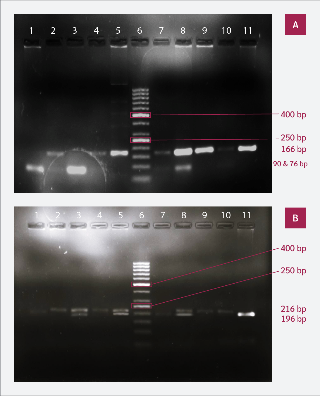
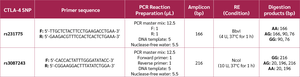
Polymerase chain reaction-restriction fragment length polymorphism (PCR-RFLP) was utilized for genotyping. The primer sequences were adopted from the methodology of Dursun et al. (18). The thermal cycling protocol consisted of an initial denaturation step at 95°C for 2 minutes, followed by 35 cycles of denaturation at 94°C, annealing at 55°C, and extension at 72°C, each lasting 30 seconds, followed by a final extension step at 72°C for 7 minutes. The PCR products were subjected to 2% agarose gel electrophoresis to verify the expected amplicon sizes (Figure 1).
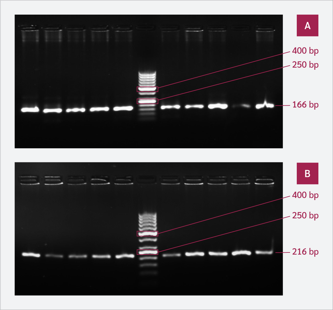
The PCR-amplified products of rs231775 and rs3087243 were digested with the restriction enzymes BbvI and NcoI (Enzynomics, Daejeon, South Korea), respectively, according to the manufacturer’s protocol. The primer sequences, amplicon sizes, corresponding restriction enzymes, and the possible genotypes determined by restriction digestion are summarized in Table 1. The digested products were resolved by electrophoresis on a 2.5% agarose gel, and the resulting bands were visualized using the Azure 600 Gel Imaging System (Azure Biosystems, Dublin, CA, USA) (Figure 2). Samples showing borderline or unclear results were re-genotyped. Additionally, to ensure reproducibility, 20 randomly selected samples were subjected to repeat genotyping with complete concordance between repeated results.
Measurement of Serum sCTLA-4 Levels
Serum sCTLA-4 levels were measured using a human sCTLA-4 enzyme-linked immunosorbent assay (ELISA) kit (Bioassay Technology Laboratory, Shanghai, China) following the manufacturer’s instructions. This kit employs a quantitative sandwich ELISA technique for the detection of circulating sCTLA-4 levels in serum samples. Standards were assayed in duplicate, 50 μL of standards and 40 μL of samples were added to the corresponding wells. Then, 10 μL of anti-sCTLA-4 antibody was added to sample wells only, followed by the addition of 50 μL of streptavidin-horseradish peroxidase (HRP) to both the sample and standard wells but not the blank well. The plate was covered with a seal and incubated for 60 minutes at 37°C. The plate was washed according to the manufacturer’s instructions, and then 50 μL of each substrate solution (A and B) was added to all wells, followed by incubation for 10 minutes in the dark at 37°C. A stop solution was then added to all wells, resulting in a color change from blue to yellow. The optical density (OD) of each well was measured using a microplate reader set to 450 nm. The concentrations were determined from a standard curve and expressed in ng/mL.
Statistical Analysis
Statistical analyses were performed using the Statistical Package for the Social Sciences (SPSS), version 25.0 (IBM Corp., Armonk, NY, USA). Non-parametric quantitative variables were summarized as medians and interquartile ranges (IQRs), while qualitative variables were presented as frequencies and percentages.
Non-parametric continuous variables were analyzed using the Mann-Whitney U test for two-group comparisons and the Kruskal-Wallis test for multiple-group comparisons. The relationships among categorical variables were evaluated using the chi-square test or the Monte Carlo test when appropriate.
Spearman’s rank correlation coefficient was utilized to determine the association between quantitative variables. HaploView software version 4.2 (Broad Institute, Cambridge, MA, USA) was used to estimate haplotype frequencies. To account for multiple comparisons, the Benjamini-Hochberg false discovery rate (FDR) correction was applied separately to case-control analyses and within-case clinical association analyses. Both raw p-values and adjusted p-values (q-values) were reported. Statistical significance was defined using an adjusted p<0.05.
Results
The present case-control study enrolled 45 patients with Pv and 45 age- and sex-matched healthy controls. The median age of the patients with Pv was 40 years, with an IQR of 26.0–50.0, while the median age of the controls was 36 years, with an IQR of 24.0–46.0. The Pv group included 26 males and 19 females, whereas the control group included 21 males and 24 females.
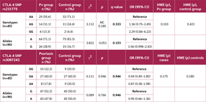
Table 2:
Distribution of genotypes and alleles for the studied SNPs among the Pv and control groups.
Regarding genotype distribution, both study groups were in Hardy-Weinberg equilibrium (HWE) for both SNPs. Furthermore, no significant differences in genotype or allele frequencies were detected between the two groups for either SNP (Table 2).
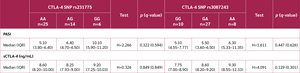
Table 3 illustrates the associations between the studied SNPs genotypes and the examined parameters within the Pv group, with no significant differences observed.

Four haplotypes defined by rs231775 and rs3087243 SNPs were identified, with the G/G haplotype occurring significantly more frequently in the Pv group (Table 4).

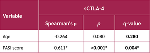
Regarding the sCTLA-4 immunoassay, serum sCTLA-4 levels differed significantly between the Pv group and controls (Table 5). Notably, serum sCTLA-4 levels correlated positively with PASI scores but not with age (Table 6). Moreover, there was no significant difference in sCTLA-4 levels by sex within the Pv group, with a median (IQR) of 8.60 (8.05–9.80) ng/mL in males and 8.40 (7.75–9.60) ng/mL in females (U=218.0, p=0.504, q=0.588).
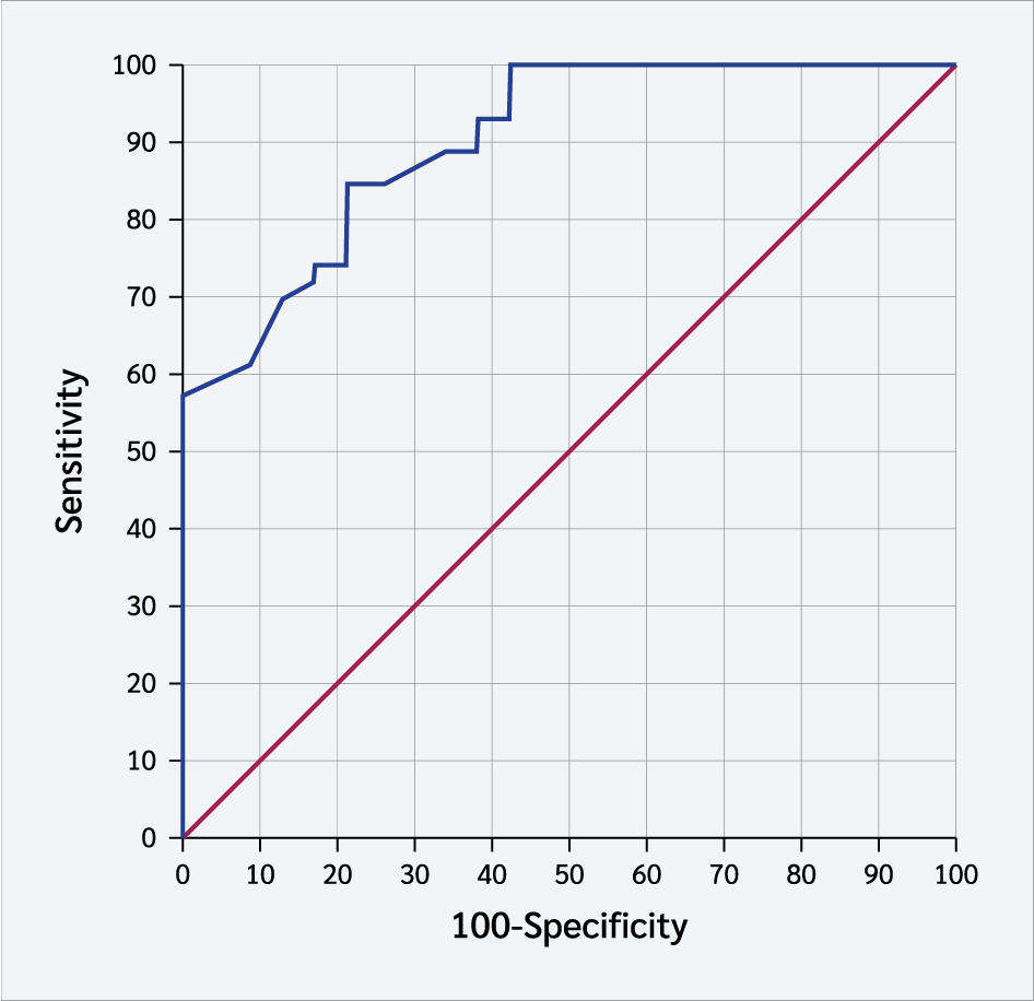
The receiver operating characteristic (ROC) analysis revealed excellent discriminatory performance of serum sCTLA-4 levels in differentiating the Pv group from controls, with an area under the curve (AUC) of 0.914. The optimal cutoff value was ≥ 7.35 ng/mL, yielding a sensitivity of 86.67%, specificity of 80%, and an overall accuracy of 83.3% (Figure 3).
Discussion
the CTLA-4 gene, it was essential to investigate the relevance of CTLA-4 polymorphisms to Pv susceptibility in the Egyptian population. This study examined the relationship between two CTLA-4 SNPs (rs231775 and rs3087243) and serum sCTLA-4 levels and Pv susceptibility in Egyptian adults.
Our study revealed a male-to-female ratio of 1.37:1 in the Pv group. This reflects a slight male predominance, consistent with previous studies from Egypt (19), Saudi Arabia (20), and the Maghreb region (21), which reported ratios of 1.2:1, 1.4:1, and 1.16:1, respectively. However, this difference may be coincidental and could be attributed to a higher number of male patients attending the clinic or a higher willingness among male patients to participate in the study.
For rs231775, neither genotype nor allele frequencies differed significantly within the Pv group. Although the G allele showed a higher frequency among patients with Pv, this difference did not reach statistical significance (p=0.051), and this borderline value was not maintained after correction for multiple testing (adjusted p-value=0.153). Our findings align with studies in Korean, Japanese, and Caucasian populations reporting no association with Pv (22-25).
In disagreement with our findings, Dursun et al. (18) reported a strong association between this SNP and Pv, identifying the G allele as a potential risk factor.
Similar to the findings of Dursun et al. (18) and Łuszczek et al. (25), we found no correlation between rs231775 SNP genotypes and disease severity. Additionally, our results demonstrated no association between this SNP and sCTLA-4 levels in the serum of patients with Pv, aligning with the findings of Berry et al. (17) and Huang et al. (26). In contrast, a study by Gallardo et al. (27) identified a potential association between this SNP and sCTLA-4 expression, with GG genotype carriers exhibiting reduced serum sCTLA-4 levels. Notably, rs231775 is located within an exon, rather than an intron, promoter region, or the 3′ untranslated region (3′-UTR), which are more directly involved in transcription and mRNA splicing (28,29). This makes it less likely that the rs231775 SNP directly influences sCTLA-4 levels. One possible explanation for the conflicting findings is that rs231775 may be in strong linkage disequilibrium with other nearby regulatory SNPs that do affect sCTLA-4 expression.
Similarly, no significant difference was observed in the frequency of rs3087243 genotypes between the Pv and control groups, consistent with the findings of Łuszczek et al. (25) but contrasting with Dursun et al. (18), who reported significantly higher frequencies of the GG genotype and G allele in cases. However, consistent with Łuszczek et al. (25) and Dursun et al. (18), we found no relationship between rs3087243 genotypes and disease severity.
Several factors may explain the conflicting genotypic association findings, including the relatively small sample size of our study. Additionally, autoimmune diseases are multifactorial, influenced by interactions between genetic, immunological, and environmental factors, and variation in ethnic background across populations may also affect the impact of specific variants on different diseases (30-32).
The rs3087243 SNP is hypothesized to influence splicing efficiency and the production of sCTLA-4 mRNA isoforms. According to Ueda et al. (33), the G allele was reported to result in decreased sCTLA-4 mRNA expression and consequently decreased serum sCTLA-4 levels. However, we observed no correlation between sCTLA-4 levels and different rs3087243 genotypes, consistent with reports by Berry et al. (17), López et al. (34) and Dahal et al. (35), which similarly found that this SNP was not associated with sCTLA-4 levels in autoimmune diseases.
This discrepancy may be attributed to differences in the methodologies used. In 2003, Ueda et al. (33) measured sCTLA-4 mRNA levels rather than the circulating serum protein levels. Assays based on ELISA may not differentiate between the sCTLA-4 proteins expressed from sCTLA-4 mRNA and other CTLA-4 immunoreactive forms, including those generated by proteolytic processing of membrane-bound CTLA-4 (mCTLA-4) (36). Additionally, elevated serum sCTLA-4 may reflect reduced protein turnover rather than increased gene expression, and lower mRNA levels could be the result of feedback regulatory mechanisms (17).
Haplotype analysis identified four haplotypes defined by rs231775 and rs3087243. The G/G haplotype was significantly more common in the Pv group, suggesting a potential synergistic effect. This contradicts Łuszczek et al. (25), who found no significant differences in haplotype distribution among the studied population. Comparisons with Dursun et al. (18) are challenging, as their study included an additional variant in their three-SNP analysis.
Assessment of serum sCTLA-4 revealed significantly higher levels in patients with Pv than in controls. Although ROC analysis demonstrated a strong discriminatory performance between the Pv and control groups, psoriasis vulgaris remains a clinical diagnosis, and the current findings suggest only a preliminary role for sCTLA-4 as a supportive biomarker. Furthermore, its potential clinical utility may be limited by the observation that elevated sCTLA-4 levels have also been reported in other autoimmune diseases (37). To our knowledge, this is the first study to investigate sCTLA-4 as a potential biomarker in psoriasis. Furthermore, sCTLA-4 levels correlated positively with disease severity, but showed no association with age or sex among the participants.
In agreement with our findings, Łuszczek et al. (24) also reported a significantly higher mean concentration in serum of patients with Pv, particularly in patients with higher PASI scores. Contrary to our findings, Liu et al. (38) described a lack of correlation between sCTLA-4 levels and psoriasis severity. However, in their study, severity was assessed using categorical groups (mild, moderate, and severe) rather than PASI scores as a continuous variable, as applied in our analysis.
These findings may suggest a role for sCTLA-4 in the immunopathogenesis of Pv. Previous studies have proposed that sCTLA-4 modulates immune responses by acting as a decoy receptor that competes with CD28 for B7 binding, thereby disrupting activation through the B7/CD28 co-stimulatory pathway (39-41). Experimental evidence further suggests that the biological effects of sCTLA-4 are context-dependent and may be dual in nature. While sCTLA-4 may inhibit the activation of resting T cells by limiting CD28-mediated co-stimulation, in activated immune environments, it may also compete with mCTLA-4 for B7 ligands, thereby preventing mCTLA-4 engagement and the delivery of its intracellular inhibitory signals. Consequently, the immunoregulatory functions mediated by mCTLA-4, including suppression of IL-2 production and IL-2 receptor expression, inhibition of the expression of transcription factors, and attenuation of APC activation through reduced IL-12 production, may be diminished, favoring persistent T-cell activation and inflammation (24,42). Moreover, disruption of co-stimulatory pathways by CTLA-4 Ig analogs has been shown to promote Th17 polarization, which is a vital driver of psoriasis pathogenesis (43).
Our study has several limitations, including the small sample size and the single-center design, which may have limited the statistical power to detect weaker associations and the generalizability of the results. Additionally, sequencing-based validation and gene expression analysis were not performed due to financial constraints. Moreover, complete blinding of laboratory personnel to the clinical status of enrolled participants was not feasible.
Future studies should include larger, multicenter cohorts. We recommend investigating additional SNPs and conducting functional assays to clarify the biological effects of CTLA-4 polymorphisms. Moreover, examining the relationship between sCTLA-4 mRNA expression and serum protein levels may provide insight into potential regulatory mechanisms. Longitudinal studies are also recommended to evaluate changes in sCTLA-4 levels over time and their association with disease activity.
Conclusion
This study provides new insights into the association of CTLA-4 SNPs rs231775 and rs3087243 with psoriasis vulgaris. Although individual SNPs were not linked to disease risk, the G/G haplotype showed a significant association, suggesting a possible synergistic effect. Moreover, elevated sCTLA-4 may contribute to immune dysregulation, promoting disease severity by interfering with mCTLA-4 signaling and facilitating a shift toward Th17-driven inflammation in psoriasis vulgaris.
Ethical Approval
The study protocol was approved by the Ethical Committee of the Faculty of Medicine, Ain Shams University on June 14, 2023 with decision number FMASU MS 334/2023.
Informed Consent
Written informed consent was obtained from all participants before enrollment in the study.
Peer-review
Externally peer-reviewed
Author Contributions
Concept – N.H.K., I.H.S.; Design – N.H.K.; Supervision – F.E.Y.F., I.H.S.; Funding – N.H.K.; Materials – N.H.K.; Data Collection and/or Processing – F.E.Y.F., M.A.S., N.H.K.; Analysis and/or Interpretation – F.E.Y.F., M.A.S., N.H.K.; Literature Review – N.H.K.; Writing – N.H.K., M.A.S.; Critical Review – I.H.S., F.E.Y.F.
Conflict of Interest
The authors declared no conflict of interest.
Financial Disclosure:
The authors declared that this study has received no financial support.
AI Statement
The authors used ChatGPT to assist with grammar and language refinement in selected sections. All AI-assisted sections were carefully reviewed and revised by the authors, who take full responsibility for the accuracy of the submitted content.
References
Yan F, Tao J, Liu J, Chen Y, Huang Z. Cross-tissue transcriptome-wide association study reveals novel psoriasis susceptibility genes. J Transl Autoimmun. 2025;10:100286. [CrossRef]
Skayem C, Taieb C, Halioua B, Baissac C, Saint Aroman M. Epidemiology of psoriasis: a worldwide global study. Acta Derm Venereol. 2025;105:adv42945. [CrossRef]
Global Psoriasis Atlas. Prevalence heat map [Internet]. Manchester: Global Psoriasis Atlas. [cited April 1, 2026]. Available from: https://www.globalpsoriasisatlas.org/en/explore/prevalence-heatmap
World Health Organization. Global report on psoriasis [Internet]. Geneva: World Health Organization; 2016. [cited April 1, 2026]. Available from: https://www.who.int/publications/i/item/9789241565189
Chiricozzi A, Romanelli P, Volpe E, Borsellino G, Romanelli M. Scanning the immunopathogenesis of psoriasis. Int J Mol Sci. 2018;19(1):179. [CrossRef]
Hu P, Wang M, Gao H, Zheng A, Li J, Mu D, et al. The role of helper T cells in psoriasis. Front Immunol. 2021;12:788940. [CrossRef]
Deshmukh SK. CTLA-4 and PD-L1/PD-1 pathways: immune checkpoint inhibitors and cancer immunotherapy. J Cancer Immunol. 2020;2(1):10–2. [CrossRef]
Salinas-Santander MA, Suárez-Valencia VJ, Ángel-Martínez MD, Kubelis-Lopez DE, Zapata-Salazar NA, Ocampo-Garza JA, et al. Association between the CTLA4 +49A/G (rs231775) and CT60 (rs3087243) gene variants with vitiligo: study on a Mexican population. An Bras Dermatol. 2022;97(6):710–5. [CrossRef]
Rallis KS, Hillyar CRT, Sideris M, Davies JK. T-cell-based immunotherapies for haematological cancers, Part A: A SWOT Analysis of Immune Checkpoint Inhibitors (ICIs) and Bispecific T-Cell Engagers (BiTEs). Anticancer Res. 2021;41(3):1123–41. [CrossRef]
Hosseini A, Gharibi T, Marofi F, Babaloo Z, Baradaran B. CTLA-4: from mechanism to autoimmune therapy. Int Immunopharmacol. 2020;80:106221. [CrossRef]
van de Kerkhof PC. From Empirical to pathogenesis-based treatments for psoriasis. J Invest Dermatol. 2022;142(7):1778–85. [CrossRef]
Yao Y, Zeng L, Huang X, Zhang J, Zhang G, Wang L. Role of coinhibitory molecules in the treatment of psoriasis (Review). Exp Ther Med. 2024;27(5):209. [CrossRef]
Sharma C, R Ali B, Osman W, Afandi B, Aburawi EH, Beshyah SA, et al. Association of variants in PTPN22, CTLA-4, IL2-RA, and INS genes with type 1 diabetes in Emiratis. Ann Hum Genet. 2021;85(2):48–57. [CrossRef]
Elshazli R, Settin A, Salama A. Cytotoxic T lymphocyte associated antigen-4 (CTLA-4) +49 A>G gene polymorphism in Egyptian cases with rheumatoid arthritis. Gene. 2015;558(1):103–7. [CrossRef]
Shehjar F, Dil-Afroze, Misgar RA, Malik SA, Laway BA. A significant association of the CTLA4 gene variants with the risk of autoimmune Graves' disease in ethnic Kashmiri population. Cell Immunol. 2020;347:103995. [CrossRef]
National Center for Biotechnology Information (US). dbSNP: rs3087243 [Internet]. Bethesda (MD): National Library of Medicine (US); [cited April 1, 2026]. Available from: https://www.ncbi.nlm.nih.gov/snp/rs3087243
Berry A, Tector M, Oaks MK. Lack of association between sCTLA-4 levels in human plasma and common CTLA-4 polymorphisms. J Negat Results Biomed. 2008;7:8. [CrossRef]
Dursun HG, Yılmaz HO, Dursun R, Kulaksızoğlu S. Association of cytotoxic T lymphocyte antigen-4 gene polymorphisms with psoriasis vulgaris: a case-control study in Turkish population. J Immunol Res. 2018;2018:1643906. [CrossRef]
El-Komy MHM, Mashaly H, Sayed KS, Hafez V, El-Mesidy MS, Said ER, et al. Clinical and epidemiologic features of psoriasis patients in an Egyptian medical center. JAAD Int. 2020;1(2):81–90. [CrossRef]
Fatani MI, Abdulghani MH, Al-Afif KA. Psoriasis in the eastern Saudi Arabia. Saudi Med J. 2002;23(2):213–7. [CrossRef]
Mikrani J, Shrestha A. Clinical and epidemiological features of psoriasis in patients visiting Lumbini Medical College. J Lumbini Med Coll. 2014;2(1):1–3. [CrossRef]
Kim YK, Pyo CW, Hur SS, Kim TY, Kim TG. No associations of CTLA-4 and ICAM-1 polymorphisms with psoriasis in the Korean population. J Dermatol Sci. 2003;33(1):75–7. [CrossRef]
Tsunemi Y, Saeki H, Kishimoto M, Mitsui H, Tada Y, Torii H, et al. Cytotoxic T lymphocyte antigen-4 gene (CTLA4) polymorphisms in Japanese patients with psoriasis vulgaris. J Dermatol Sci. 2003;32(2):163–5. [CrossRef]
Łuszczek W, Kubicka W, Jasek M, Baran E, Cisło M, Nockowski P, et al. CTLA-4 gene polymorphisms and natural soluble CTLA-4 protein in psoriasis vulgaris. Int J Immunogenet. 2006;33(3):217–24. [CrossRef]
Łuszczek W, Majorczyk E, Nockowski P, Pluciński P, Jasek M, Nowak I, et al. Distribution of the CTLA-4 single nucleotide polymorphisms CT60G>A and +49A>G in psoriasis vulgaris patients and control individuals from a Polish Caucasian population. Int J Immunogenet. 2008;35(1):51–5. [CrossRef]
Huang X, Liu X, Ye Y, Zhang T, Mei S, Zhu T, et al. Polymorphisms and circulating plasma protein levels of immune checkpoints (CTLA-4 and PD-1) are associated with Posner-Schlossman syndrome in Southern Chinese. Front Immunol. 2021;12:607966. [CrossRef]
Gallardo D, Bosch-Vizcaya A, Rodríguez-Romanos R, Santos N, Buño I, de la Cámara R, et al; GvHD/Immunotherapy Committee of the Spanish Hematopoietic Transplant Group. Donor CTLA-4 genotype modulates the immune response to minor histocompatibility antigen mismatches. Biol Blood Marrow Transplant. 2017;23(12):2042–7. [CrossRef]
Wang XB, Zhao X, Giscombe R, Lefvert AK. A CTLA-4 gene polymorphism at position -318 in the promoter region affects the expression of protein. Genes Immun. 2002;3(4):233–4. [CrossRef]
Deng N, Zhou H, Fan H, Yuan Y. Single nucleotide polymorphisms and cancer susceptibility. Oncotarget. 2017; 8(66):110635–49. [CrossRef]
Sirota M, Schaub MA, Batzoglou S, Robinson WH, Butte AJ. Autoimmune disease classification by inverse association with SNP alleles. PLoS Genet. 2009;5(12):e1000792. [CrossRef]
Gyu Song G, Ho Lee Y. CTLA-4 +49A/G and -318C/T polymorphisms and susceptibility to multiple sclerosis: a meta-analysis. Immunol Invest. 2013;42(5):409–22. [CrossRef]
Lin HY, Huang PY, Tseng TS, Park JY. SNPxE: SNP-environment interaction pattern identifier. BMC Bioinformatics. 2021;22(1):425. [CrossRef]
Ueda H, Howson JM, Esposito L, Heward J, Snook H, Chamberlain G, et al. Association of the T-cell regulatory gene CTLA4 with susceptibility to autoimmune disease. Nature. 2003;423(6939):506–11. [CrossRef]
López-Villalobos EF, Carrillo-Ballesteros FJ, Muñoz-Valle JF, Palafox-Sánchez CA, Valle Y, Orozco-Barocio G, et al. Association of CD28 and CTLA4 haplotypes with susceptibility to primary Sjögren's syndrome in Mexican population. J Clin Lab Anal. 2019;33(1):e22620. [CrossRef]
Dahal LN, Barker RN, Ward FJ. The soluble isoform of CTLA-4 correlates with interferon-α activity in systemic lupus erythematosus. J Rheumatol. 2020;47(2):302–4. [CrossRef]
Ward FJ, Dahal LN, Wijesekera SK, Abdul-Jawad SK, Kaewarpai T, Xu H, et al. The soluble isoform of CTLA-4 as a regulator of T-cell responses. Eur J Immunol. 2013;43(5):1274–85. [CrossRef]
Malkawi AK, Nimer RM, Almogren M, Masood A, Alarfaj AS, Benabdelkamel H, et al. Quantitative analysis of soluble costimulatory molecules as potential diagnostic biomarkers for rheumatoid arthritis using LC-MS/MS in MRM mode. Clin Chim Acta. 2023;548:117501. [CrossRef]
Liu P, He Y, Wang H, Kuang Y, Chen W, Li J, et al. The expression of mCTLA-4 in skin lesion inversely correlates with the severity of psoriasis. J Dermatol Sci. 2018;89(3):233–40. [CrossRef]
Kristiansen OP, Larsen ZM, Pociot F. CTLA-4 in autoimmune diseases – a general susceptibility gene to autoimmunity? Genes Immun. 2000;1(3):170–84. [CrossRef]
Aprilia A, Handono K, Sujuti H, Sabarudin A, Winaris N. sCD163, sCD28, sCD80, and sCTLA-4 as soluble marker candidates for detecting immunosenescence. Immun Ageing. 2024;21(1):9. [CrossRef]
Gerold KD, Zheng P, Rainbow DB, Zernecke A, Wicker LS, Kissler S. The soluble CTLA-4 splice variant protects from type 1 diabetes and potentiates regulatory T-cell function. Diabetes. 2011;60(7):1955–63. [CrossRef]
Saverino D, Simone R, Bagnasco M, Pesce G. The soluble CTLA-4 receptor and its role in autoimmune diseases: an update. Auto Immun Highlights. 2010;1(2):73–81. [CrossRef]
Bouguermouh S, Fortin G, Baba N, Rubio M, Sarfati M. CD28 co-stimulation down regulates Th17 development. PLoS One. 2009;4(3):e5087. [CrossRef]
VOLUME
,
ISSUE
Correspondence
Received
Accepted
Published
Suggested Citation
DOI
License

