
Anti-ADAMTSL5 and Anti-K17 Autoantibodies in Psoriasis: Diagnostic Performance and Associations with Systemic Inflammation


Abstract
Objective:
Psoriasis is a chronic inflammatory skin disease that can mimic several common dermatoses, highlighting the need for objective diagnostic biomarkers. This study aimed to evaluate the diagnostic utility of serum anti-ADAMTSL5 and anti-K17 autoantibodies in psoriasis and to assess their associations with systemic inflammation.
Materials and Methods:
In this case-control study, complete blood count (CBC) parameters were assessed in patients with psoriasis and healthy controls using an automated hematology analyzer, and serum anti-ADAMTSL5 and anti-K17 antibody levels were measured by enzyme-linked immunosorbent assay (ELISA). Diagnostic accuracy and associations between variables were assessed using receiver operating characteristic (ROC) curve and univariable linear regression analyses.
Results:
The study enrolled 120 participants, including 80 patients with psoriasis and 40 healthy controls. Total leukocyte and absolute neutrophil counts were significantly higher in patients with psoriasis than in healthy controls (p<0.001). Serum levels of anti-ADAMTSL5 (2.18 ± 0.64 ng/mL vs. 0.06 ± 0.04 ng/mL) and anti-K17 (13.74 ± 2.78 ng/L vs. 6.28 ± 0.83 ng/L) were significantly higher in patients with psoriasis (p=0.000). Univariable regression analyses showed that both autoantibodies were significantly associated with the systemic neutrophilic burden. Moreover, both biomarkers demonstrated high diagnostic performance on ROC analysis, with an area under the curve (AUC) of 1.000 for anti-ADAMTSL5 and 0.998 for anti-K17.
Conclusion:
Serum anti-ADAMTSL5 and anti-K17 antibodies demonstrated promising diagnostic potential for distinguishing patients with psoriasis from healthy controls in the present study. Given their strong associations with neutrophilic leukocytosis, these findings suggest a potential association between cutaneous autoimmunity and systemic inflammation.
Keywords:
Psoriasis biomarkers autoantibodies anti-ADAMTSL5 anti-K17Introduction
Psoriasis is a chronic immune-mediated inflammatory skin disease that can be challenging to distinguish from clinically similar dermatoses (1). Early-stage psoriasis may be confused with atopic dermatitis, nummular eczema, and pityriasis rosea. This diagnostic uncertainty can lead to a delay in diagnosis, which is important because timely treatment may help reduce the risk of systemic comorbidities, including psoriatic arthritis and metabolic syndrome. Thus, serological biomarkers are needed to help distinguish psoriasis from its clinical mimics (2).
One major autoantigen implicated in psoriasis is ADAMTSL5, a melanocyte-derived protein recognized by autoreactive CD8+ T cells (3). ADAMTSL5 is a large glycoprotein expressed by melanocytes in the epidermis. In genetically susceptible individuals, particularly those carrying HLA-C*06:02, ADAMTSL5-derived peptides can be presented to CD8+ T cells, triggering an autoimmune response that contributes to localized inflammation and tissue damage at the epidermal-melanocyte interface (4).
In healthy epidermis, keratin 17 (K17) expression is very low; however, inflammatory cytokines in psoriatic lesions markedly induce its expression in keratinocytes (5). Increased K17 expression and keratinocyte damage may enhance the exposure and presentation of K17-derived peptides to the adaptive immune system. In genetically susceptible individuals, loss of self-tolerance to these peptides can promote the activation of autoreactive T cells and subsequently B-cell activation and production of anti-K17 autoantibodies (6). Moreover, molecular mimicry between K17-derived peptides and streptococcal M proteins may contribute to cross-reactive immune responses, providing a possible link between streptococcal infection and psoriasis exacerbation (7). ADAMTSL5- and K17-associated immune responses have both been implicated in inflammatory pathways relevant to psoriasis, particularly the interleukin-23/interleukin-17 (IL-23/IL-17) axis, which promotes keratinocyte hyperproliferation and sustains chronic inflammation. ADAMTSL5 represents a melanocyte-associated autoantigen capable of activating autoreactive T-cell responses, whereas increased K17 expression in stressed keratinocytes may contribute to continued antigenic stimulation and inflammatory amplification (8).
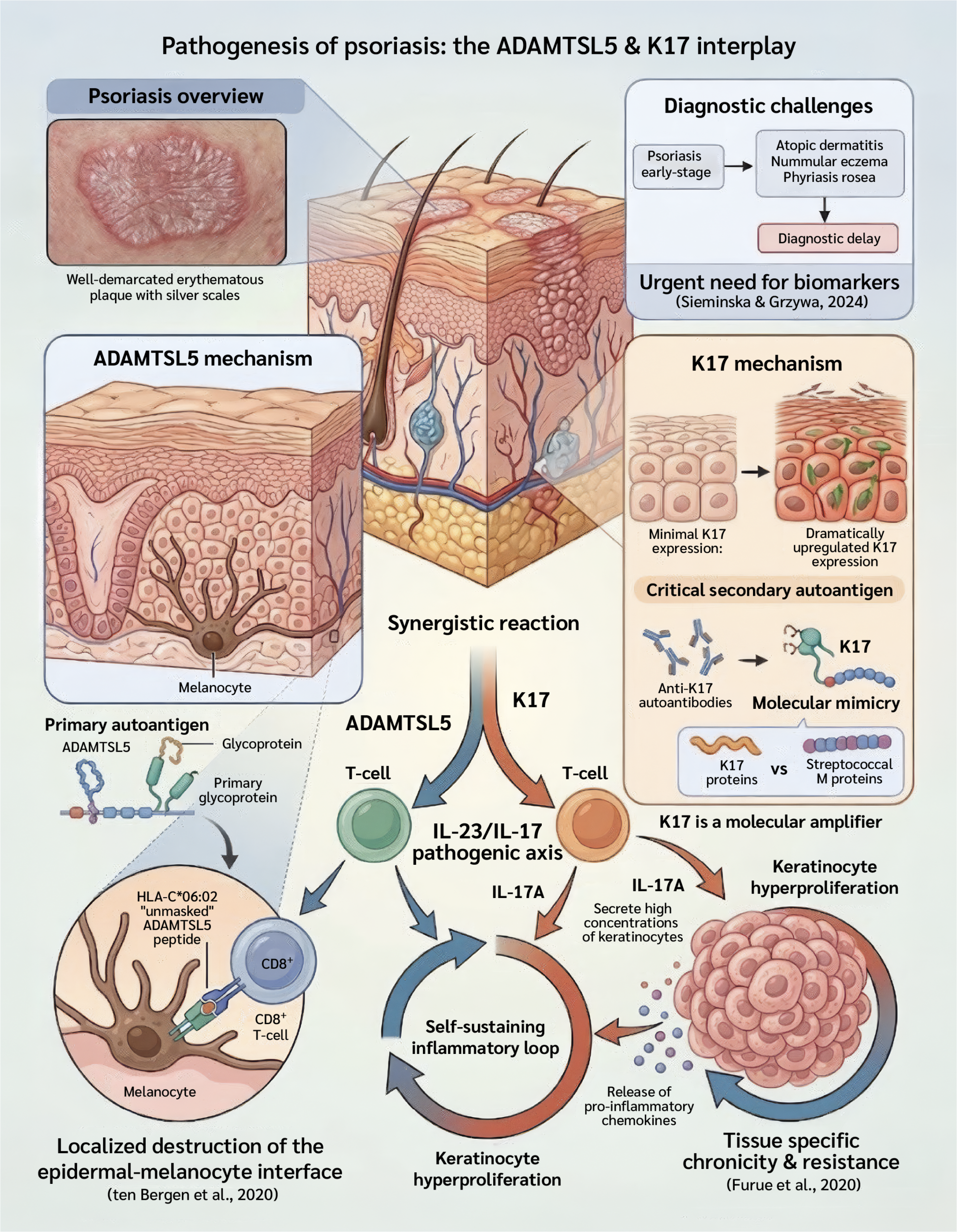
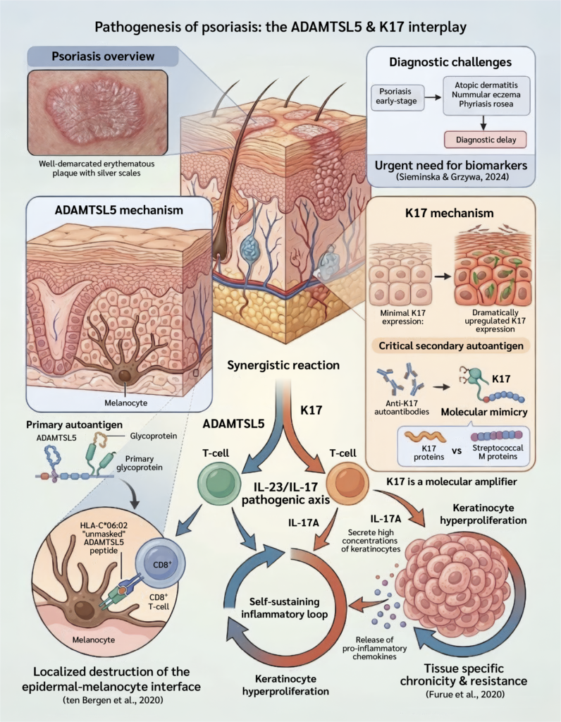
Based on these previously described mechanisms, we propose that ADAMTSL5- and K17-associated immune responses may represent interconnected components of a feed-forward inflammatory framework rather than a formally established direct interaction between the two autoantigens. This conceptual framework is summarized in Figure 1.
The objective of this study was to assess systemic hematological changes by comparing complete blood count (CBC) profiles, with particular emphasis on total white blood cell (WBC), neutrophil, lymphocyte, and monocyte counts. We also investigated whether circulating anti-ADAMTSL5 and anti-K17 autoantibody levels were associated with systemic inflammatory parameters, particularly the circulating neutrophil count. Therefore, the current study aimed to evaluate the individual diagnostic performance of serum anti-ADAMTSL5 and anti-K17 autoantibodies and to investigate their statistical associations with systemic inflammatory parameters. The concurrent involvement in psoriasis was interpreted within the proposed inflammatory framework and was not evaluated as a combined diagnostic model.
Materials and Methods
Samples were collected from patients with psoriasis under the supervision of a specialist dermatologist at İbn Sina Teaching Hospital in Mosul, Iraq, between December 1, 2025, and March 1, 2026. Scientific analyses were conducted in the specialized laboratories of the Department of Biology, College of Science, University of Mosul.
Adult participants were included in the final analysis, comprising patients with psoriasis and healthy controls. Psoriasis was clinically diagnosed and confirmed by a consultant dermatologist. Pediatric participants identified during the initial recruitment period were excluded from the final study population and statistical analyses. Eligible participants were adults aged 18–65 years with a clinically confirmed diagnosis of psoriasis. Patients with autoimmune diseases, acute or chronic infections, liver or kidney disorders, malignancies, and hormonal disorders were excluded. Participants receiving immunosuppressive medication, biological therapy, or systemic corticosteroids were also excluded, as were pregnant or breastfeeding women.
Venous blood (5 mL) was drawn from each participant into two tubes: 2 mL into an ethylenediaminetetraacetic acid (EDTA) tube for complete blood count (CBC) analysis and 3 mL in a gel tube for serum separation by centrifugation at 1500 rpm for 5 min. The separated serum was then collected and stored for subsequent analysis.
Hematological Analysis
Complete blood count (CBC) and five-part white blood cell differential (5-DIFF) analyses were performed using an automated hematology analyzer (Mindray BC-5000, Shenzhen, China). Whole blood samples collected in tubes containing dipotassium EDTA were analyzed (9).
Measurement of Anti-ADAMTSL5 and Anti-K17 Autoantibodies
Serum anti-ADAMTSL5 and anti-K17 autoantibody levels were quantitatively determined using commercial indirect enzyme-linked immunosorbent assay (ELISA) kits according to the manufacturer's instructions: anti-ADAMTSL5 (SunLong Biotech Co., Ltd., catalog no. SL4833Hu, Hangzhou, China) and anti-K17 (SunLong Biotech Co., Ltd., catalog no. SL0202Hu, Hangzhou, China). The assays were performed using microtiter plates precoated with the corresponding recombinant human antigens. Standard solutions were prepared by serial dilution to obtain concentrations of 18, 12, 6, 3, and 1.5 ng/mL, and standards were analyzed in duplicate. Serum samples were diluted 1:5 by mixing 10 µL of serum with 40 µL of sample diluent. Then, 50 µL of each prepared standard or diluted serum sample was added to the designated antigen-coated wells, with one well serving as a blank control.
The plates were sealed and incubated at 37°C for 30 min, after which the wells were aspirated and washed five times with diluted wash buffer. Following washing, 50 µL of horseradish peroxidase (HRP)-conjugated detection reagent was added to each well, except the blank, and the plates were incubated at 37°C for an additional 30 min. The washing procedure was then repeated five times to remove unbound conjugate.
For color development, 50 µL of chromogen solution A followed by 50 µL of chromogen solution B was added to each well. The plates were gently mixed and incubated at 37°C for 15 min in the dark. The enzymatic reaction was terminated by adding 50 µL of stop solution, resulting in a color change from blue to yellow. Optical density was measured at 450 nm using a BioTek microplate reader within 15 min of adding the stop solution, with the blank well used for baseline correction. Autoantibody concentrations were calculated from the corresponding standard curves generated using the known standard concentrations (10).
Statistical Analysis
Data were analyzed using IBM SPSS Statistics version 24.0 (IBM Corp., Armonk, NY, USA). The normality of the data distribution was evaluated using the Shapiro-Wilk test. All continuous variables, including leukocyte and differential counts, met the assumption of normality (p>0.05). Therefore, parametric statistical tests were applied, and data are expressed as the mean ± standard deviation (SD) and range. Independent-samples t tests and one-way analysis of variance (ANOVA) were used for group comparisons after assessing homogeneity of variance. Receiver operating characteristic (ROC) curve analysis, including the area under the curve (AUC) and 95% confidence interval (CI), was used to evaluate the diagnostic performance of anti-ADAMTSL5 and anti-K17 antibodies. Univariable linear regression models were used to evaluate the individual statistical associations of each autoantibody level with neutrophil counts. A two-tailed p<0.05 was considered statistically significant (11).
Results
The final analysis included 120 adult participants: 80 patients with psoriasis (35 males and 45 females) and 40 healthy controls (21 males and 19 females).
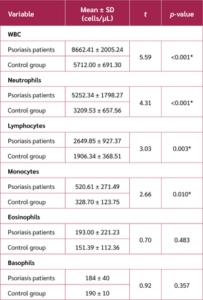
The hematological profile demonstrated significant systemic variations between patients with psoriasis and healthy controls. Total WBC counts were significantly higher in the psoriasis group than in the control group. Analysis of the five-part differential showed significant increases in absolute neutrophil, lymphocyte, and monocyte counts. Conversely, basophil (BAS) and eosinophil (EOS) counts did not differ significantly between the two groups (Table 1).
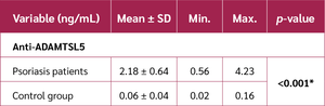
Serum anti-ADAMTSL5 concentrations were significantly higher in patients with psoriasis than in healthy controls. As shown in Table 2, patients with psoriasis had markedly higher autoantibody levels (2.18 ± 0.64 ng/mL) than healthy controls (0.06 ± 0.04 ng/mL; p<0.001). This finding indicates a significant difference in serum anti-ADAMTSL5 levels between the two groups.
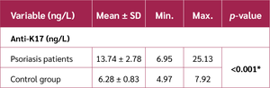
Serum anti-K17 concentrations were also significantly higher in patients with psoriasis than in healthy controls. As detailed in Table 3, patients with psoriasis had significantly higher autoantibody levels (13.74 ± 2.78 ng/L) than healthy controls (6.28 ± 0.83 ng/L; p<0.001). This finding indicates a significant difference in serum anti-K17 levels between the two groups.

A univariable linear regression analysis was performed to evaluate the association between serum anti-ADAMTSL5 levels and neutrophil counts. As shown in Table 4, anti-ADAMTSL5 levels were positively associated with neutrophil counts (B=720.89, p=0.042). The model explained approximately 10% of the observed variation in neutrophil counts (R²=0.10), as seen in Table 4.

A univariable linear regression analysis was performed to evaluate the association between anti-K17 levels and neutrophil counts. As shown in Table 5, anti-K17 levels were positively associated with neutrophil counts (B=217.94, p=0.007). The model explained approximately 11% of the observed variation in neutrophil counts (R²=0.11), as seen in Table 5.

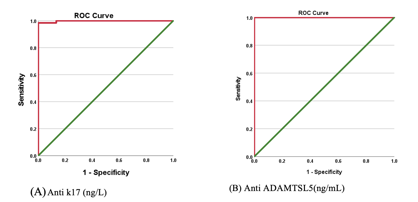
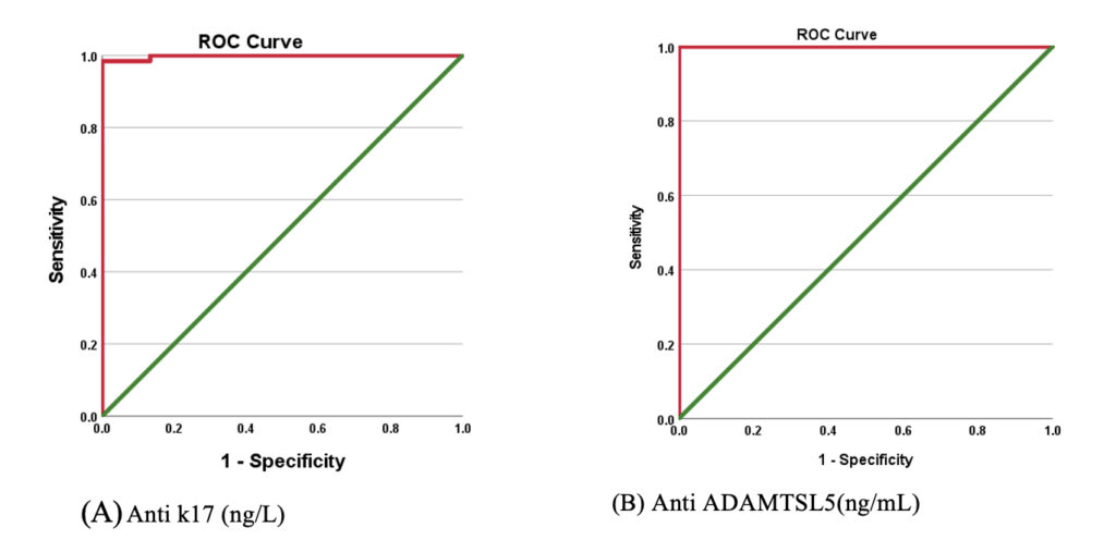
The diagnostic performance of anti-ADAMTSL5 and anti-K17 was evaluated using ROC curve analysis. As shown in Table 6, both biomarkers demonstrated excellent discrimination between patients with psoriasis and healthy controls. Anti-ADAMTSL5 achieved an AUC of 1.000, whereas anti-K17 showed an AUC of 0.998, indicating excellent diagnostic performance in this study population.
Discussion
In our study, we demonstrated significantly elevated total WBC counts in patients with psoriasis compared with healthy controls, with the increase primarily attributable to higher neutrophil counts. This finding is consistent with the chronic inflammatory nature of psoriasis and the established involvement of neutrophils in Munro microabscess formation and amplification of T helper 17 (Th17)/T helper 22 (Th22)-mediated responses (12). Lymphocyte and monocyte counts were also significantly higher in patients with psoriasis, supporting the involvement of both adaptive and innate immune responses. In contrast, eosinophil and basophil counts were not significantly different between the groups, consistent with the predominance of Th17-mediated rather than Th2-mediated inflammation in psoriasis (13).
We found that serum anti-ADAMTSL5 levels were markedly elevated in patients with psoriasis compared with healthy controls. ADAMTSL5 is a melanocyte-derived autoantigen presented in the context of HLA-C*06:02 and recognized by autoreactive CD8+ T cells, providing a link between melanocyte-specific autoimmunity and psoriatic inflammation (14). The increased anti-ADAMTSL5 levels observed in our study are consistent with its proposed role as a psoriasis-associated autoantigen and may reflect persistent immune activation within the psoriatic microenvironment (15,16). The relationship between this autoimmune response and the IL-23/IL-17 inflammatory axis may also contribute to leukocyte accumulation and keratinocyte hyperproliferation (17).
Similarly, serum anti-K17 concentrations were significantly higher in patients with psoriasis than in healthy controls. K17 is an inducible intermediate filament protein that functions as a stress-associated signal and is upregulated following epidermal injury and exposure to inflammatory cytokines, including IL-17 and interferon-gamma (IFN-γ) (18). Autoimmune recognition of K17 may therefore reflect loss of immune tolerance within the chronic inflammatory environment. Previous studies have linked K17 to T-cell activation, neutrophil recruitment, and maintenance of the inflammatory response (19,20). These findings further support the potential utility of anti-K17 as a serological marker of psoriasis-associated immune dysregulation.
Univariable linear regression analysis demonstrated a statistically significant positive association between serum anti-ADAMTSL5 levels and neutrophil counts. However, this association should not be interpreted as evidence of a direct causal effect of anti-ADAMTSL5 on neutrophil expansion. Rather, the parallel elevation of anti-ADAMTSL5 and neutrophils may reflect concurrent activation of autoimmune and innate inflammatory pathways within the systemic inflammatory environment of psoriasis. ADAMTSL5-specific autoimmunity and neutrophilic inflammation may therefore represent interconnected manifestations of the underlying inflammatory process, with neutrophil recruitment additionally mediated by chemokines such as C-X-C motif chemokine ligand 1 (CXCL1) and C-X-C motif chemokine ligand 8 (CXCL8) (21).
In a separate univariable linear regression model, serum anti-K17 levels were also positively associated with neutrophil counts. K17 is upregulated in psoriatic epidermis and participates in inflammatory pathways involving IL-17/IL-23 signaling and neutrophil-recruiting chemokines such as C-C motif chemokine ligand 2 (CCL2), CXCL1, and CXCL8 (22). Nevertheless, the relatively low R² indicates that anti-K17 accounts for only a limited proportion of the variation in neutrophil counts, emphasizing the multifactorial nature of neutrophilic inflammation in psoriasis (23,24).
The diagnostic performance of both biomarkers was evaluated using ROC analysis and was high in this study population. Anti-ADAMTSL5 showed an AUC of 1.000, while anti-K17 showed an AUC of 0.998, indicating excellent discrimination between patients with psoriasis and healthy controls (25,26). The corresponding cutoff values further support their potential utility as serological biomarkers. However, these findings should be interpreted within the context of the present study population and require validation in larger independent cohorts including patients with other inflammatory and clinically similar dermatological conditions before their broader diagnostic application can be established (27,28).
Conclusion
Our study demonstrated significantly elevated serum anti-ADAMTSL5 and anti-K17 levels in patients with psoriasis compared with healthy controls. The diagnostic performance of both biomarkers was high based on ROC analysis. These findings support the potential of anti-ADAMTSL5 and anti-K17 as adjunctive serological biomarkers for psoriasis, particularly in clinical settings where psoriasis may overlap morphologically with inflammatory dermatoses such as atopic dermatitis, nummular eczema, and pityriasis rosea. However, given the relatively limited sample size and case-control design, validation in larger independent cohorts that directly include these clinical mimickers is required before their differential diagnostic utility can be established.
The significant association between autoantibody levels and systemic hematological manifestations, particularly neutrophilic leukocytosis, suggests that these biomarkers may reflect not only local but also systemic autoimmune inflammation.
Overall, anti-ADAMTSL5 and anti-K17 showed strong individual diagnostic performance and were positively associated with systemic inflammatory features. Their potential complementary diagnostic value warrants further evaluation using combined multivariable models and independent validation cohorts.
Ethical Approval
This study was approved by the Scientific and Ethical Committee of the Department of Life Sciences, College of Sciences, University of Mosul on November 9, 2025 (Approval no. 50619). Ethical approval for the collection of patient information was also obtained under the supervision of the Ministry of Health in accordance with the Declaration of Helsinki.
Informed Consent
Not applicable.
Peer-review
Externally peer-reviewed
Author Contributions
Concept – D.A.M.A., R.G.M.A.; Design – D.A.M.A., R.G.M.A.; Supervision – R.G.M.A.; Funding – D.A.M.A., R.G.M.A.; Materials – D.A.M.A., R.G.M.A.; Data Collection and/or Processing – D.A.M.A., R.G.M.A.; Analysis and/or Interpretation – D.A.M.A., R.G.M.A.; Literature Review – D.A.M.A., R.G.M.A.; Writing – D.A.M.A.; Critical Review – R.G.M.A.; Other – Graphic Abstract, D.A.M.A., R.G.M.A.
Conflict of Interest
The authors declared that they have no conflict of interest.
Financial Disclosure:
The authors declared that this study has received no financial support.
AI Statement
The authors declared that no artificial intelligence (AI) tools or generative AI technologies were used in the writing, editing, or preparation of this manuscript.
Acknowledgement
The authors would like to express their sincere gratitude to Dr. Rojan G. M. Al-Allaf, Department of Biology, College of Science, for providing technical and academic support during the completion of this study. The authors also thank Amin Abd Elaziz, Al Sultan Lab., for their encouragement and cooperation in completing the manuscript.
References
Sieminska I, Pieniawska M, Grzywa TM. The immunology of psoriasis-current concepts in pathogenesis. Clin Rev Allergy Immunol. 2024;66(2):164–91. [CrossRef]
Zhou X, Zhou H, Luo X, Wu RF. Discovery of biomarkers in the psoriasis through machine learning and dynamic immune infiltration in three types of skin lesions. Front Immunol. 2024;15:1388690. [CrossRef]
Ten Bergen LL, Petrovic A, Aarebrot AK, Appel S. Current knowledge on autoantigens and autoantibodies in psoriasis. Scand J Immunol. 2020;92(4):e12945. [CrossRef]
Anand S, Littler DR, Mobbs JI, Braun A, Baker DG, Tennant L, et al. Complimentary electrostatics dominate T-cell receptor binding to a psoriasis-associated peptide antigen presented by human leukocyte antigen C*06:02. J Biol Chem. 2023;299(7):104930. [CrossRef]
Lin Y, Zhang W, Li B, Wang G. Keratin 17 in psoriasis: Current understanding and future perspectives. Semin Cell Dev Biol. 2022;128:112–9. [CrossRef]
Luo Y, Pang B, Hao J, Li Q, Qiao P, Zhang C, et al. Keratin 17 covalently binds to alpha-enolase and exacerbates proliferation of keratinocytes in psoriasis. Int J Biol Sci. 2023;19(11):3395–411.[CrossRef]
Liu T, Li S, Ying S, Tang S, Ding Y, Li Y, et al. The IL-23/IL-17 pathway in inflammatory skin diseases: from bench to bedside. Front Immunol. 2020;11:594735. [CrossRef]
Furue M, Furue K, Tsuji G, Nakahara T. Interleukin-17A and keratinocytes in psoriasis. Int J Mol Sci. 2020;21(4):1275. [CrossRef]
Xiang D, Yue J, Lan Y, Sha C, Ren S, Li Y, et al. Evaluation of Mindray BC-5000 hematology analyzer: a new miniature 5-part WBC differential instrument. Int J Lab Hematol. 2015;37(5):597–605. [CrossRef]
Al-Heyali H, Al-Allaf R. Novelty in colorectal cancer biomarkers: the predictive value and clinical utility of the carcinoembryonic antigen and aldehyde dehydrogenase 1B1 autoantibodies for assessing tumour biology and the cancer stem cell burden. Asian Pac J Cancer Biol. 2025;10(4):905–13.[CrossRef]
Emir B, Can FE, Kaymaz E, Ozel Z, Efgan MG, Tekindal MA, et al. Sample size and power analysis for ROC AUC differences in diagnostic tests: a methodological evaluation of the Obuchowski-McClish and Hanley-McNeil methods. BMC Med Res Methodol. 2026;26(1):43. [CrossRef]
Matsushima Y, Hwang ST, Simon SI. Mechanobiology of neutrophil inflammasome signaling in psoriasis. Curr Opin Immunol. 2025;97:102680. [CrossRef]
Armstrong AW, Read C. Pathophysiology, Clinical presentation, and treatment of psoriasis: a review. JAMA. 2020;323(19):1945–60. [CrossRef]
Li L, Lu J, Liu J, Wu J, Zhang X, Meng Y, et al. Immune cells in the epithelial immune microenvironment of psoriasis: emerging therapeutic targets. Front Immunol. 2024;14:1340677. [CrossRef]
Favaro R, Facheris P, Formai A, Gargiulo L, Ibba L, Fiorillo G, et al. Autoreactivity to self-antigens LL37 and ADAMTSL5 influences the clinical response to risankizumab in psoriatic patients. J Autoimmun. 2024;147:103244. [CrossRef]
Brembilla NC, Boehncke WH. Revisiting the interleukin 17 family of cytokines in psoriasis: pathogenesis and potential targets for innovative therapies. Front Immunol. 2023;14:1186455. [CrossRef]
Andersen CSB, Kvist-Hansen A, Siewertsen M, Enevold C, Hansen PR, Kaur-Knudsen D, et al. Blood cell biomarkers of inflammation and cytokine levels as predictors of response to biologics in patients with psoriasis. Int J Mol Sci. 2023;24(7):6111. [CrossRef]
Romashin DD, Tolstova TV, Varshaver AM, Kozhin PM, Rusanov AL, Luzgina NG. Keratins 6, 16, and 17 in health and disease: a summary of recent findings. Curr Issues Mol Biol. 2024;46(8):8627–41. [CrossRef]
Xu J, Jin L, Zhang T, Zhuang Y, Zhu Z, Liu Y, et al. Keratin 17 enhances the chemotaxis of neutrophils through regulating CXCL1 secretion in keratinocytes via PI3K/AKT/NF-κB pathway in psoriasis. Chin Med J (Engl). 2026;139(5):782–4. Erratum in: Chin Med J (Engl). 2026;139(14):2175. [CrossRef]
Albanesi C, Madonna S, Gisondi P, Girolomoni G. The interplay between keratinocytes and immune cells in the pathogenesis of psoriasis. Front Immunol. 2018;9:1549. [CrossRef]
Fan X, Shu P, Wang Y, Ji N, Zhang D. Interactions between neutrophils and T-helper 17 cells. Front Immunol. 2023;14:1279837. [CrossRef]
Chen HL, Lo CH, Huang CC, Lu MP, Hu PY, Chen CS, et al. Galectin-7 downregulation in lesional keratinocytes contributes to enhanced IL-17A signaling and skin pathology in psoriasis. J Clin Invest. 2021;131(1):e130740. [CrossRef]
Gupta RK, Gracias DT, Figueroa DS, Miki H, Miller J, Fung K, et al. TWEAK functions with TNF and IL-17 on keratinocytes and is a potential target for psoriasis therapy. Sci Immunol. 2021;6(65):eabi8823. [CrossRef]
Kar BR, Sathishkumar D, Tahiliani S, Parthasarathi A, Neema S, Ganguly S, et al. Biomarkers in psoriasis: the future of personalised treatment. Indian J Dermatol. 2024;69(3):256–63. [CrossRef]
Martín Monreal MT, Kvist-Hansen A, Massarenti L, Steffensen R, Loft N, Hansen PR, et al. Characterization of circulating extracellular traps and immune responses to citrullinated LL37 in psoriasis. Front Immunol. 2023;14:1247592. [CrossRef]
Wang L, Liu R, Tang Y, Ma Y, Wang G, Ruan Q, et al. Advances in psoriasis research: decoding immune circuits and developing novel therapies. Int J Mol Sci. 2025;26(18):9233. [CrossRef]
Ishimoto T, Arakawa Y, Vural S, Stöhr J, Vollmer S, Galinski A, et al. Multiple environmental antigens may trigger autoimmunity in psoriasis through T-cell receptor polyspecificity. Front Immunol. 2024;15:1374581. [CrossRef]
Zhu J, Shi XF, Chu CQ. Autoantibodies in psoriatic arthritis: are they of pathogenic relevance? Chin Med J (Engl). 2020;133(24):2899–901. [CrossRef]
VOLUME
,
ISSUE
Correspondence
Received
Accepted
Published
Suggested Citation
DOI
License

