
Immune-Biochemical Stratification Identifies the Pure Primary Biliary Cholangitis Phenotype in AMA-Positive Individuals


Abstract
Objective:
The presence of antimitochondrial antibodies (AMAs) is strongly associated with primary biliary cholangitis (PBC), but serological reactivity alone may not consistently characterize a distinct clinical presentation. We aimed to determine whether a combination of mitochondrial-specific autoimmunity (M2 seropositivity) and cholestatic burden, quantified by alkaline phosphatase (ALP), is associated with a physiologically coherent pure PBC phenotype within an AMA-positive cohort.
Materials and Methods:
This retrospective cross-sectional study included 300 AMA-positive adult patients assessed at a tertiary referral institution. Clinical phenotypes were categorized as pure PBC, PBC-autoimmune hepatitis (AIH) overlap, or non-PBC based on accepted diagnostic criteria.
Results:
The study included 300 AMA-positive individuals, of which 80 were diagnosed with pure PBC, 37 with PBC-AIH overlap, and 183 were categorized as non-PBC. Seropositivity for M2 and ALP levels were independently associated with the pure PBC phenotype. M2 positivity was strongly associated with pure PBC (adjusted odds ratio [aOR] 14.60, 95% confidence interval [CI] 1.56–136.37, p=0.019), whereas each 10 U/L increment in ALP was associated with 18% higher odds of pure PBC (aOR 1.18, 95% CI 1.05–1.32, p=0.005). The multivariable model showed good internal discrimination (area under the receiver operating characteristic curve [AUC] 0.86, 95% CI 0.76–0.93), achieving 69.0% sensitivity and 89.2% specificity at the optimal probability threshold. In sensitivity analyses involving overlap syndromes, ALP remained independently correlated with disease phenotype, whereas the influence of M2 seropositivity diminished.
Conclusion:
Antimitochondrial antibody positivity alone does not define a consistent clinical phenotype. The integration of M2 seropositivity with ALP levels may provide a more informative immune-biochemical framework for phenotypic stratification among AMA-positive individuals.
Keywords:
Primary, biliary, cholangitis antimitochondrial, antibody M2, seropositivity alkaline, phosphatase cholestasis autoimmunityIntroduction
Primary biliary cholangitis (PBC) is a chronic immune-mediated cholestatic liver disease marked by progressive destruction of small intrahepatic bile ducts, potentially leading to fibrosis and cirrhosis if left untreated (1,3). Historically considered an organ-specific autoimmune disorder, contemporary evidence indicates a multidimensional immunopathogenic framework in which adaptive autoimmunity, innate immune activation, cholangiocyte-intrinsic stress responses, and bile acid-mediated epithelial injury collectively contribute to chronic biliary inflammation (1-3). Genome-wide association studies have identified susceptibility loci within human leukocyte antigen (HLA) class II and immune regulatory pathways, reinforcing the concept of systemic immunological dysregulation as an underlying determinant of disease risk (1,2). The significant female predominance and frequent association with other autoimmune diseases further suggest a common immunogenetic and immunoregulatory basis (1).
The serological marker of PBC is the development of antimitochondrial antibodies (AMAs), specifically those directed against the E2 subunit of the pyruvate dehydrogenase complex (AMA-M2), which are present in around 90–95% of individuals with confirmed disease (4,5). Mitochondrial-directed autoimmunity constitutes a highly distinctive humoral signature of autoimmune liver disease, indicating a selective breach of tolerance to highly conserved intracellular antigens (5,6). Cholangiocyte apoptosis, abnormal antigen presentation, and bile acid-induced epithelial stress are mechanistically suggested to reveal immunogenic mitochondrial epitopes, thus sustaining autoreactive B- and T-cell responses (3,6).
Although AMA and M2 antibodies are highly specific for established PBC, serological reactivity alone does not reliably identify disease manifestation or progression. Community-based studies have shown that AMA positivity occurs in approximately 0.2–0.4% of the general population, although anti-M2 positivity is significantly rarer (7). Large observational and longitudinal studies have shown that only a small proportion of seropositive individuals without early cholestasis develop clinically evident PBC during follow-up (8-10). These findings demonstrate a clear distinction between autoimmune reactivity and clinically significant cholangiopathy, implying that additional immunological or biochemical signals are required to characterize clinically relevant phenotypes.
In addition to standard AMA responses, disease-specific antinuclear antibodies, such as anti-gp210 and anti-sp100, have been associated with distinct clinical subgroups and, in rare cases, more severe disease outcomes (5,11). From a biological perspective, portal infiltration by autoreactive CD4+ and CD8+ T cells, T helper 1 (Th1)– and T helper 17 (Th17)–driven immunological polarization, and dysregulated cytokine signaling all contribute to biliary epithelial damage (1,6). Recent studies have increasingly suggested that innate immune activation and cholangiocyte-specific inflammatory signaling pathways are critical to the breakdown of immune tolerance (6). Collectively, these findings support a multidimensional immunologic hypothesis in which serological markers constitute only one aspect of disease biology.
However, despite progress in understanding disease mechanisms, a significant translational gap persists. In AMA-positive populations, the relationship between mitochondrial-specific autoimmunity and measurable biochemical damage in identifying clinically significant disease phenotypes remains unclear. In clinical practice, AMA-positive individuals without persistent cholestasis frequently constitute a diagnostic gray area, while overlap syndromes further obscure phenotypic distinctions (8,12). A systematic framework that integrates immunological specificity with the biochemical manifestation of biliary damage may improve phenotypic resolution.
The objective of this study was to investigate whether the combination of mitochondrial-specific autoimmunity (M2 seropositivity) and cholestatic burden, measured by alkaline phosphatase (ALP) levels, can differentiate the pure PBC phenotype within an AMA-positive population. We hypothesized that an integrated immune–biochemical framework would be associated with improved phenotypic stratification compared with AMA status alone and would better reflect an immunologic gradient among AMA-positive individuals.
Materials and Methods
This retrospective cross-sectional study included consecutive AMA-positive adult patients identified through the Medical Microbiology Laboratory of Ankara Bilkent City Hospital between February 2019 and July 2023. Antimitochondrial antibody and antinuclear antibody (ANA) indirect immunofluorescence (IIF) intensity grades, ANA staining patterns, AMA-M2 immunoblot results, and clinical diagnoses were retrospectively reviewed.
All consecutive adult patients (≥18 years) with confirmed AMA positivity were eligible for inclusion. Antimitochondrial antibody positivity was established based on the laboratory-specific cutoff levels determined by the IIF assay. Patients were required to have available data on M2 serostatus and baseline biochemical parameters, including ALP, to be included in the final analysis.
Patients with active viral hepatitis (positive hepatitis B virus [HBV] DNA, hepatitis C virus [HCV] RNA, or acute hepatitis E), decompensated cirrhosis (Child-Pugh class B/C, active ascites, variceal bleeding, or hepatic encephalopathy), active malignancy, active cholestasis (including biliary obstruction on ultrasonography, magnetic resonance cholangiopancreatography [MRCP], or computed tomography [CT]; choledocholithiasis; biliary malignancy; or primary sclerosing cholangitis) or insufficient clinical, serologic, or biochemical data for multivariable analysis were excluded. These criteria were applied to ensure that identified immune-biochemical relationships accurately represented intrinsic autoimmune characteristics while minimizing confounding from secondary cholestatic or inflammatory liver injury.
Clinical phenotypes were determined according to established international diagnostic criteria and multidisciplinary specialist evaluation. Patients were categorized as pure PBC, PBC-autoimmune hepatitis (AIH) overlap, or non-PBC within the AMA-positive cohort based on clinical, serologic, and biochemical findings at the time of assessment.
Pure PBC was defined as the presence of AMA and persistent cholestatic liver biochemistry, particularly elevated ALP, without significant hepatitic inflammatory features. The diagnosis was established according to internationally accepted diagnostic criteria and confirmed by the attending hepatologists.
The PBC-AIH overlap syndrome was defined by the coexistence of diagnostic features of both PBC and AIH. These features included elevated transaminases, increased immunoglobulin G (IgG) levels, and consistent histopathological findings when available. The diagnosis was established using accepted clinical criteria, including the Paris-type criteria, and multidisciplinary specialist consensus. Patients who did not meet the criteria for either pure PBC or PBC-AIH overlap were categorized as non-PBC. All diagnoses were confirmed by experienced hepatologists and gastroenterologists during the clinical evaluation.
Hospital records were reviewed to obtain data on alanine aminotransferase (ALT), aspartate aminotransferase (AST), ALP, gamma-glutamyl transferase (GGT), international normalized ratio (INR), albumin, immunoglobulin M (IgM), and IgG. Laboratory parameters were assessed either simultaneously with ANA testing or within one week thereafter. Furthermore, the records included clinical details such as age, sex, and diagnosis.
An ANA IIF test was performed using HEp-20-10/liver tissues (Euroimmun AG, Lübeck, Germany). The screening technique began with a preliminary dilution of 1:100 as recommended by the manufacturer. At the screening dilution of 1:100, fluorescence intensity was graded semiquantitatively as 0 (negative), 1+, 2+, 3+, or 4+, with 4+ representing the strongest fluorescence intensity using a EUROSTAR III Plus fluorescent microscope (Euroimmun AG, Lübeck, Germany) at ×40 magnification. The presence of antibodies against extractable nuclear antigens (ENA) was assessed using the EUROLINE ANA Profile 3 plus DFS70 (IgG) test kit manufactured by manufactured by Euroimmun AG, Lübeck, Germany.
The study was conducted in compliance with the Declaration of Helsinki and was authorized by the Ankara Bilkent City Hospital Non-Interventional Clinical Research Ethics Committee on July 12, 2023, with decision number E2-23-4480.
Statistical Analysis
R version 4.3.2 (R Foundation for Statistical Computing, Vienna, Austria) and IBM SPSS Statistics for Windows, version 22.0 (IBM Corp., Armonk, NY, USA) were used for statistical analyses. The Shapiro-Wilk test and visual examination of histograms were used to determine whether continuous variables were normally distributed. Most continuous variables were presented as medians (interquartile range [IQR]) because they were not normally distributed. Categorical variables were expressed as frequencies and percentages.
The Kruskal-Wallis test for continuous variables and the chi-square test or Fisher's exact test for categorical variables, as applicable, were used to compare the three clinical groups (pure PBC, overlap, and non-PBC).
A multivariable logistic regression model was used to identify independent predictors of the pure PBC phenotype based on characteristics that were deemed clinically significant or showed a potential association in univariate analysis. Odds ratios (ORs) with 95% confidence intervals (CIs) were used to present the results. Multicollinearity among predictor variables was assessed using the variance inflation factor (VIF) analysis before multivariable logistic regression modeling. Variables with VIF values below 5 were considered to have no significant multicollinearity.
Receiver operating characteristic (ROC) curve analysis based on the predicted probabilities from the adjusted multivariable logistic regression model was performed to evaluate discriminative performance. The area under the curve (AUC) was calculated to quantify model performance. The Youden index was used to determine the optimal probability threshold, which was then applied to calculate the model's sensitivity and specificity. A two-sided p<0.05 was considered statistically significant.
Results
From February 2019 to July 2023, 35,195 autoimmune liver antibody panel tests (ANA, anti-smooth muscle antibody [ASMA], and liver-kidney microsomal [LKM] antibody) were performed at our institution. Of these tests, 1009 were positive for antimitochondrial antibodies (2.9%). Additionally, 40,297 M2 immunoblot tests were performed, of which 1349 were positive (3.3%). The study included a total of 300 patients who tested positive for AMA and underwent M2 immunoblot testing. The mean age of the included patients was 57.0 (48.0–66.0) years.
Primary biliary cholangitis was diagnosed in 80 patients, and PBC-AIH overlap in 37 (12.3%). Among the remaining 183 patients, 46 (25.1%) had rheumatologic diseases (ankylosing spondylitis, Behçet disease, mixed connective tissue disease, myositis, psoriatic arthritis, Sjögren syndrome, Raynaud phenomenon, scleroderma, systemic lupus erythematosus, and adult-onset Still disease); 45 (24.6%) had neurologic diseases (Alzheimer disease, Bell's palsy, epilepsy, tension-type headache, migraine, multiple sclerosis, hemiplegia, myopathy, neuropathic bladder, polyneuropathy, cerebrovascular accident, vertigo, and optic neuritis); 30 (16.4%) had endocrine diseases (diabetes mellitus, hypothyroidism, hypoparathyroidism, multinodular goiter, pituitary adenoma, and chronic thyroiditis); 18 (9.8%) had gastroenterological diseases (cryptogenic cirrhosis, pancreatitis, hepatic fibrosis, Crohn disease, nonalcoholic fatty liver disease, pancreatic cyst, pangastritis, and hepatic hemangioma); 12 (6.6%) had renal diseases (chronic renal failure and hydronephrosis); 12 (6.6%) had pulmonary diseases (asthma, chronic obstructive pulmonary disease, pneumonia, interstitial lung disease, and pulmonary arterial hypertension); 4 (2.2%) had cardiovascular diseases (hypertension, heart failure, and pericarditis); and 16 (8.7%) had other conditions (joint pain, leukocytoclastic vasculitis, immune thrombocytopenic purpura, anemia, dyspepsia, xerosis cutis, allergy, and urticaria).
The study population comprised 300 AMA-positive individuals categorized into three distinct phenotypes: pure PBC, PBC-AIH overlap, and non-PBC AMA-positive patients. A distinct serologic gradient developed among phenotypes: pure PBC (n=80, 26.7%), PBC-AIH overlap (n=37, 12.3%), and non-PBC (n=183, 61.0%). Intensity grades of AMA IIF differed significantly among the clinical phenotypes (p=0.0002), with higher grades observed in the cholestatic phenotypes.
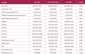
M2 positivity was more frequent in pure PBC (92.5%) and PBC-AIH overlap (75.7%) than in non-PBC patients (59.0%) (p<0.001), underscoring its phenotypic specificity among AMA-positive populations. Although overall ANA positivity rates (pure PBC, 43/80 [53.8%]; overlap, 22/37 [59.5%]; non-PBC, 84/183 [45.9%]) did not differ significantly (p=0.224), semiquantitative ANA IIF intensity grades differed modestly among the clinical phenotypes (p=0.022).
Immunoglobulin profiling showed modest but statistically significant increases in IgM levels in pure PBC patients (1.90 [1.48–2.81] g/L) compared to non-PBC patients (1.30 [1.00–2.10] g/L; overlap: 1.44 [1.04–2.67] g/L) (p=0.047), while IgG levels remained comparable (p=0.276), indicating a predominance of humoral cholestatic immune activation over generalized hypergammaglobulinemia. Liver biochemistry revealed a strong cholestatic burden in both pure primary biliary cholangitis and overlap groups. Alkaline phosphatase and GGT levels were markedly elevated in these phenotypes (ALP: pure PBC 149.0 [102.5–252.8], overlap 160.0 [126.0–261.0], non-PBC 86.0 [67.8–112.3] U/L; GGT: pure PBC 68.5 [30.8–200.8], overlap 87.0 [50.0–149.0], non-PBC 27.0 [18.0–61.0] U/L; both p<0.001), establishing a clear biochemical distinction between cholestatic and non-cholestatic AMA-positive patients. Transaminases (AST and ALT) were significantly higher in overlap patients (AST: pure PBC 33.5 [20.8–49.0], overlap 42.0 [29.0–79.0], non-PBC 22.0 [17.0–29.8] U/L; ALT: pure PBC 40.0 [21.0–52.3], overlap 45.0 [31.0–85.0], non-PBC 25.0 [19.0–34.0] U/L; p<0.001), consistent with characteristics of hepatitic inflammation. Total bilirubin levels were considerably elevated in overlap individuals (0.65 [0.50–0.80] mg/dL vs. pure PBC 0.50 [0.40–0.74] and non-PBC 0.50 [0.40–0.66] mg/dL; p=0.029), although synthetic function measures (albumin and INR) were maintained and equivalent among groups, indicating a preponderance of early to mid-stage disease in this cohort (Table 1).
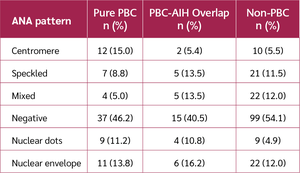
Across the entire cohort, the distribution of ANA status and patterns did not differ significantly among the clinical phenotypes (overall p=0.098). ANA negativity was the most frequent category in all three groups, while centromere, speckled, mixed, nuclear dots, and nuclear envelope patterns were distributed similarly across the phenotypes. Overall, semiquantitative ANA IIF intensity grades differed modestly among the phenotypes, whereas the distribution of qualitative ANA patterns did not differ significantly among the groups (Table 2). Collectively, these findings indicate that semiquantitative ANA IIF intensity grades differed modestly among the clinical phenotypes, whereas the distribution of qualitative ANA patterns did not differ significantly among the pure PBC, PBC-AIH overlap, and non-PBC groups (Table 2). Variance inflation factor analysis showed no evidence of problematic multicollinearity among the variables included in the multivariable logistic regression model (all VIF values <5; Supplementary Table S1).
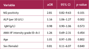
Table 3. Multivariable logistic regression analyses.
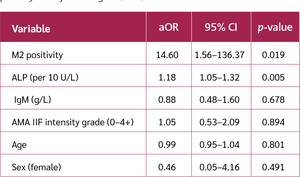
In the primary multivariable analysis excluding overlap cases, both M2 seropositivity and cholestatic burden were independently associated with the pure PBC phenotype in AMA-positive individuals. M2 positivity was strongly associated with pure PBC (adjusted OR [aOR] 14.60, 95% CI 1.56–136.37, p=0.019). The confidence interval for M2 seropositivity was wide. Alkaline phosphatase levels, quantified in 10 U/L increments, demonstrated an independent association, wherein each 10 U/L increase in ALP was associated with 18% higher odds of pure PBC (aOR 1.18, 95% CI 1.05–1.32, p=0.005). In contrast, IgM levels, AMA IIF intensity grade, age, and sex were not independently associated with pure PBC after adjustment (Table 3).
A sensitivity analysis was conducted to evaluate the robustness of these findings by defining the outcome as the PBC spectrum (pure PBC + PBC-AIH overlap) compared to non-PBC AMA-positive patients. In this comprehensive clinical definition, ALP was independently correlated with disease phenotype (aOR 1.16 per 10 U/L, 95% CI 1.06–1.27, p=0.002). Nonetheless, the effect size of M2 positivity diminished and became statistically nonsignificant (aOR 2.81, 95% CI 0.82–9.63, p=0.101), suggesting that the association of M2 seropositivity may be more specific to the pure PBC phenotype than to the broader PBC spectrum. These findings collectively suggest that among AMA-positive individuals, cholestatic biochemical burden is a consistent and broad correlate of disease phenotype, while M2 seropositivity is a more precise indicator of pure PBC (Table 3).
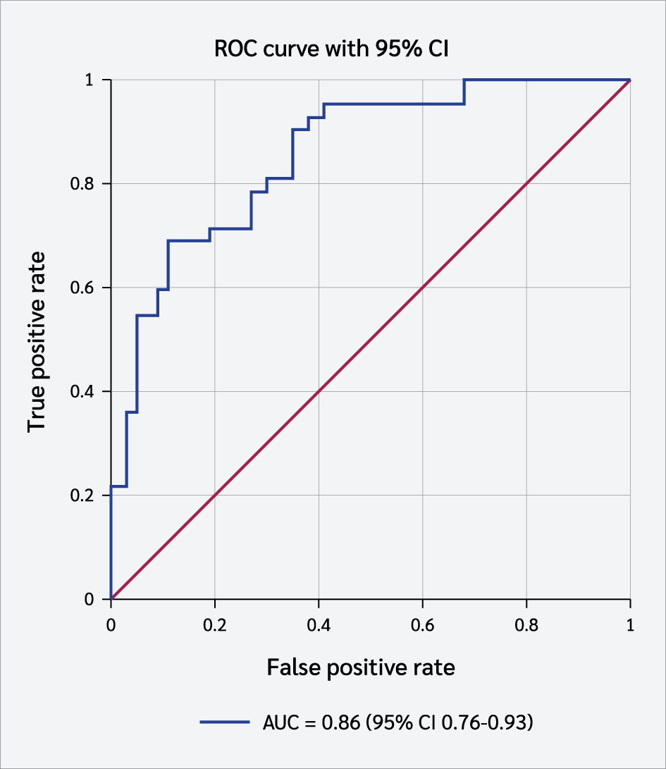
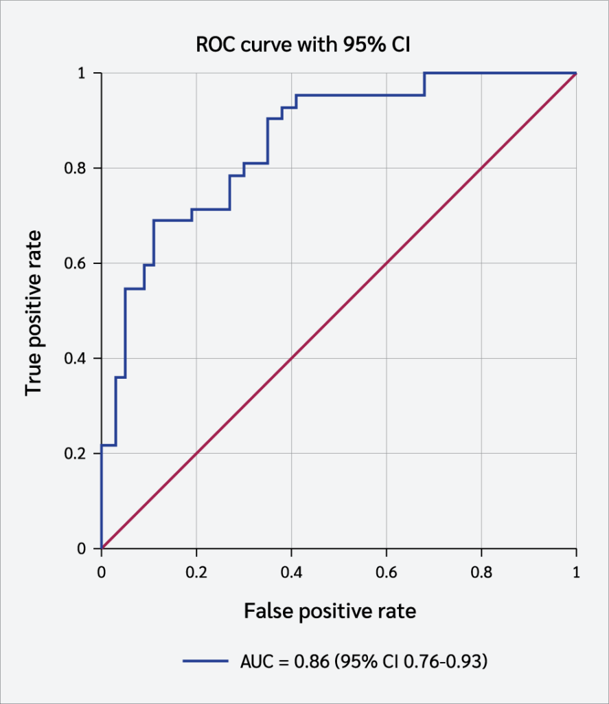
The discriminative performance of the primary multivariable model (pure PBC versus non-PBC, excluding overlap) was evaluated by receiver operating characteristic analysis. The model achieved an AUC of 0.86 (95% CI 0.76–0.93), with a sensitivity of 69.0% and a specificity of 89.2% at the optimal probability threshold (Figure 1).
Discussion
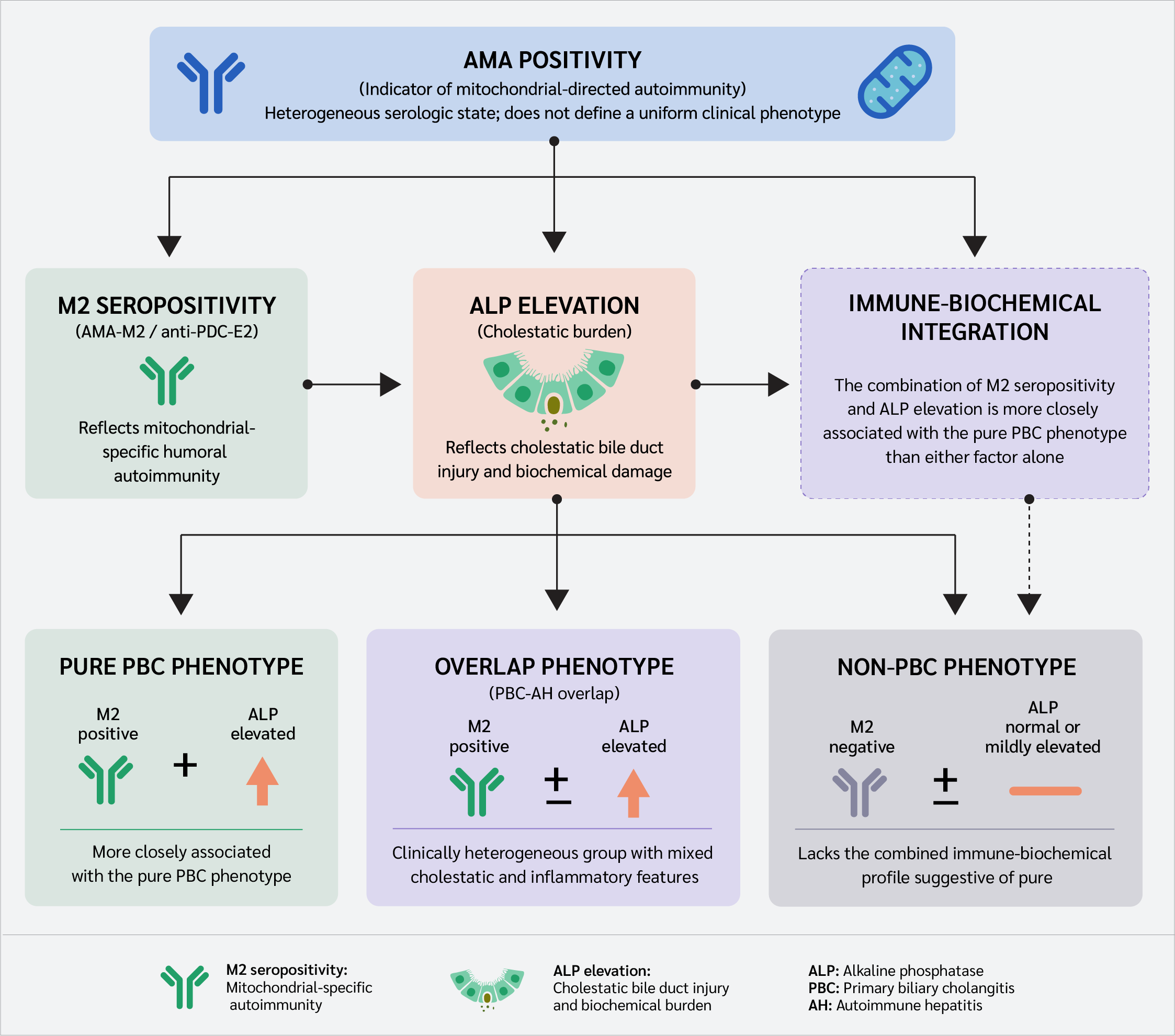
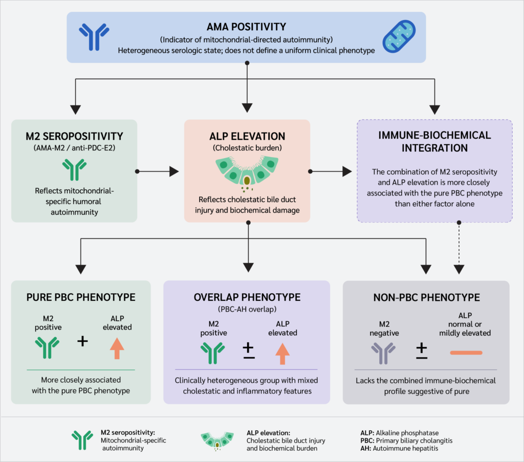
Our findings suggest that AMA positivity represents an immunologically heterogeneous state rather than a distinct clinical phenotype. Large real-world studies have shown that many AMA-positive individuals do not fulfill criteria for established PBC at baseline, indicating that serologic autoreactivity and overt cholangiopathy may dissociate in routine practice (8,10). In our cohort, AMA positivity alone did not characterize a uniform clinical phenotype. Instead, M2 seropositivity and ALP-defined cholestatic burden were associated with the pure PBC phenotype, with good internal discrimination. This integrated immune-biochemical relationship is visually summarized in our conceptual framework (Figure 2).
This finding should be interpreted cautiously. Longitudinal data show that many individuals with incidental AMA positivity, particularly those without baseline cholestasis, have a relatively low medium-term risk of progression to overt PBC (10,13). Therefore, the “pure PBC” phenotype observed cross-sectionally is not a fixed clinical entity but reflects the clinical status at evaluation. Conversely, cohorts enriched for AMA-M2 positivity may show higher rates of subsequent PBC features, suggesting that risk is heterogeneous and depends on baseline biochemical injury and ascertainment context (3).
Our hospital-based AMA and M2 prevalence rates should also be interpreted as referral-enriched estimates. Population-based studies report AMA positivity in approximately 0.2–0.4% of the general population, significantly lower than rates observed in hepatology referral cohorts (8,13). In a tertiary-care setting, pretest probability and case-mix distribution enrich the cohort for cholestatic and overlap phenotypes (14). Therefore, these estimates should not be directly generalized to unselected populations.
The association between M2 seropositivity and pure PBC is biologically plausible but should not be overinterpreted. Antimitochondrial antibodies, particularly the M2 subtype, target mitochondrial antigens, such as the E2 subunit of the pyruvate dehydrogenase complex (PDC-E2), and are closely linked to the humoral immune profile of PBC (15-18). Biliary epithelial stress and innate immune activation may enhance antigen presentation and sustain biliary-focused inflammation (6,19). However, our findings demonstrate an association rather than a causal immunopathogenic relationship. Furthermore, while our multivariable model confirmed M2 seropositivity as a strong predictor of the pure PBC phenotype, the wide confidence interval suggests that the exact magnitude of this association remains uncertain. This is likely attributable to the limited number of M2-negative cases within our specific pure PBC cohort, highlighting the need for larger, multicenter studies to more precisely define the effect size.
Although diagnostic-accuracy studies support their performance in established PBC (4,20,21), PBC-specific autoantibodies may also be detected in individuals without persistent cholestasis or in alternative conditions (22,23). This explains why M2 seropositivity alone was insufficient to characterize the broader PBC spectrum. In contrast, ALP remained independently associated with phenotype across models, functioning as a stable marker of cholestatic injury. This aligns with guidelines and outcome studies linking persistent ALP elevation to poorer transplant-free survival (3,24–27). Additionally, qualitative ANA pattern distribution did not differ significantly among clinical phenotypes (p=0.098), indicating that ANA patterns alone are insufficient to discriminate these groups in our cohort.
Overlap syndromes represent an important source of clinical heterogeneity. Criteria-based definitions, including Paris-type frameworks, are useful but may not capture the full spectrum of inflammatory cholangiopathy (28). Contemporary outcome data suggest that some clinically treated AIH-PBC variant patients respond to combination therapy without satisfying strict criteria (12). In our sensitivity analysis, the association of M2 was attenuated when overlap cases were included, whereas ALP remained significant. These findings suggest that M2 seropositivity is more closely associated with classical pure PBC, while PBC-AIH overlap syndromes represent a broader, heterogeneous inflammatory state.
From a clinical perspective, the model should be viewed as exploratory. Although the model showed good internal discrimination, its generalizability is limited without an external validation cohort. The reported AUC and probability thresholds are internally derived and may be influenced by local assay platforms or patient case-mix. Independent, multicenter validation is required before routine clinical implementation.
Individuals who are AMA-positive display profound clinical heterogeneity, ranging from asymptomatic seropositivity to evolving cholestasis or overlap syndromes. Accordingly, the observed immune–biochemical profile should be regarded as a cross-sectional representation of disease status, which may be influenced by temporal changes in liver biochemistry, immune activity, treatment exposure, and other clinical factors.
This study has several limitations. First, our single-center, retrospective design in a tertiary setting introduces selection bias, likely enriching the cohort with complex or overlapping phenotypes that do not fully reflect community-based AMA-positive populations. Second, lacking an external validation cohort, our prediction model remains exploratory. Its diagnostic metrics and probability thresholds require independent, multicenter validation before routine clinical application. Third, small sample sizes in certain subgroups, particularly ANA patterns, may have reduced statistical power. Consequently, the prevalence estimates, observed effect sizes, and model performance derived from our cohort should not be broadly generalized. For instance, the wide confidence interval observed for M2 seropositivity suggests limited precision and should be interpreted cautiously.
Finally, because of the cross-sectional design, our findings should be interpreted as associations rather than evidence of causality or disease progression. Prospective longitudinal studies are needed to determine whether AMA-positive individuals maintain their phenotype over time or transition between non-PBC, PBC-AIH overlap, and pure PBC states.
Conclusion
In conclusion, AMA positivity alone was insufficient to define a uniform clinical phenotype in this cohort. Among AMA-positive individuals, the pure PBC phenotype was more closely associated with the combined presence of M2 seropositivity and elevated ALP levels, integrating mitochondrial-specific autoimmunity with objective biochemical evidence of cholestatic injury. Compared with AMA status alone, this immune-biochemical framework may improve phenotypic stratification. However, prospective longitudinal studies and external validation are required before this model can be adopted in routine clinical practice.
Ethical Approval
The study was approved by the Ankara Bilkent City Hospital Non-Interventional Clinical Research Ethics Committee on July 12, 2023, with decision number E2-23-4480.
Informed Consent
Not applicable.
Peer-review
Externally peer-reviewed
Author Contributions
Concept – E.S.; Design – E.S.; Supervision – E.S.; Materials – E.S., S.G.; Data Collection and/or Processing – E.S., S.G.; Analysis and/or Interpretation – E.S., S.G.; Literature Review – E.S., S.G.; Writer – E.S.; Critical Reviews – E.S.
Conflict of Interest
The authors declared no conflict of interest.
Financial Disclosure:
The authors declared that this study has received no financial support.
AI Statement
During the preparation of this manuscript, the authors used an AI-based language tool (Claude, Anthropic) solely to check the grammar, language, and internal consistency of the text (e.g., cross-checking numerical values and terminology between the main text and tables). The AI tool was not used for study design, data analysis, interpretation of results, or generation of scientific content. The authors reviewed and edited all AI-assisted output and take full responsibility for the content of this publication.
References
Tanaka A, Ma X, Takahashi A, Vierling JM. Primary biliary cholangitis. Lancet. 2024;404(10457):1053–66. [CrossRef]
Lindor KD, Bowlus CL, Boyer J, Levy C, Mayo M. Primary biliary cholangitis: 2021 practice guidance update from the American Association for the Study of Liver Diseases. Hepatology. 2022;75(4):1012–3. [CrossRef]
European Association for the Study of the Liver. EASL Clinical Practice Guidelines: The diagnosis and management of patients with primary biliary cholangitis. J Hepatol. 2017;67(1):145–72. [CrossRef]
Xu Q, Zhu W, Yin Y. Diagnostic value of anti-mitochondrial antibody in patients with primary biliary cholangitis: A systemic review and meta-analysis. Medicine (Baltimore). 2023;102(45):e36039. [CrossRef]
Mimura S, Morishita A, Oura K, Yano R, Nakahara M, Tadokoro T, et al. Autoantibodies in primary biliary cholangitis: from classical markers to emerging targets. J Clin Med. 2025;14(23):8503. [CrossRef]
Chen R, Sun Y, Hu Y, Tai W. Innate immunity of bile and cholangiocytes in primary biliary cholangitis. Front Immunol. 2025;16:1655287. [CrossRef]
Manzo C, Maslinska M, Castagna A, Hysa E, Merante A, Milchert M, et al. Antimitochondrial antibodies and primary biliary cholangitis in patients with polymyalgia rheumatica/giant cell arteritis. Medicina (Kaunas). 2021;57(4):350. [CrossRef]
Dahlqvist G, Gaouar F, Carrat F, Meurisse S, Chazouillères O, Poupon R, et al; French network of Immunology Laboratories. Large-scale characterization study of patients with antimitochondrial antibodies but nonestablished primary biliary cholangitis. Hepatology. 2017;65(1):152–63. [CrossRef]
Ergenc I, Gozaydinoglu B, Keklikkiran C, Yilmaz Y. The risk of development of primary biliary cholangitis among incidental antimitochondrial M2 antibody-positive patients. Hepatol Forum. 2023;4(2):69–73. [CrossRef]
Duan W, Chen S, Li S, Lv T, Li B, Wang X, et al. The future risk of primary biliary cholangitis (PBC) is low among patients with incidental anti-mitochondrial antibodies but without baseline PBC. Hepatol Commun. 2022;6(11):3112–9. [CrossRef]
Ben Lamine Z, Ben Jazia I, Ben Ahmed M, Ben Slama A, Baccouche A, Slama F, et al. Anti-gp210 and anti-Sp100 antibodies in primary biliary cholangitis. Arab J Gastroenterol. 2021;22(4):316–20. [CrossRef]
Stoelinga AEC, Biewenga M, Drenth JPH, Verhelst X, van der Meer AJP, de Boer YS, et al; Dutch Autoimmune Hepatitis Study Group. Diagnostic criteria and long-term outcomes in AIH-PBC variant syndrome under combination therapy. JHEP Rep. 2024;6(7):101088. [CrossRef]
Metcalf JV, Mitchison HC, Palmer JM, Jones DE, Bassendine MF, James OF. Natural history of early primary biliary cirrhosis. Lancet. 1996;348(9039):1399–402. [CrossRef]
Grimes DA, Schulz KF. Bias and causal associations in observational research. Lancet. 2002;359(9302):248–52. [CrossRef]
Colapietro F, Lleo A, Generali E. Antimitochondrial antibodies: from bench to bedside. Clin Rev Allergy Immunol. 2022;63(2):166–77. [CrossRef]
Gershwin ME, Mackay IR. The causes of primary biliary cirrhosis: Convenient and inconvenient truths. Hepatology. 2008;47(2):737–45. [CrossRef]
Hirschfield GM, Dyson JK, Alexander GJM, Chapman MH, Collier J, Hübscher S, et al. The British Society of Gastroenterology/UK-PBC primary biliary cholangitis treatment and management guidelines. Gut. 2018;67(9):1568–94. [CrossRef]
Leung PS, Coppel RL, Ansari A, Munoz S, Gershwin ME. Antimitochondrial antibodies in primary biliary cirrhosis. Semin Liver Dis. 1997;17(1):61–9. [CrossRef]
Del Barrio M, Díaz-González Á, Alonso-Peña M. Primary biliary cholangitis: immunopathogenesis and the role of bile acid metabolism in disease progression. Int J Mol Sci. 2025;26(16):7905. [CrossRef]
Zhu YJ, Li J, Liu YG, Jiang Y, Cheng XJ, Han X, et al. Role of biochemical markers and autoantibodies in diagnosis of early-stage primary biliary cholangitis. World J Gastroenterol. 2023;29(34):5075–81. [CrossRef]
Hu S, Zhao F, Wang Q, Chen WX. The accuracy of the anti-mitochondrial antibody and the M2 subtype test for diagnosis of primary biliary cirrhosis: a meta-analysis. Clin Chem Lab Med. 2014;52(11):1533–42. [CrossRef]
Jallouli I, Doulberis M, Kountouras J. Primary biliary cholangitis: a summary of pathogenesis and therapies. Ann Gastroenterol. 2025;38(2):121–32. [CrossRef]
Zeng X, Lv T, Li S, Chen S, Li B, Lu Z, et al. Patients with AMA/anti-sp100/anti-gp210 positivity and cholestasis can manifest conditions beyond primary biliary cholangitis. J Clin Transl Hepatol. 2025;13(3):200–6. [CrossRef]
Lammers WJ, Hirschfield GM, Corpechot C, Nevens F, Lindor KD, Janssen HL, et al; Global PBC Study Group. Development and validation of a scoring system to predict outcomes of patients with primary biliary cirrhosis receiving ursodeoxycholic acid therapy. Gastroenterology. 2015;149(7):1804–12.e4. [CrossRef]
Carbone M, Sharp SJ, Flack S, Paximadas D, Spiess K, Adgey C, et al; UK-PBC Consortium. The UK-PBC risk scores: Derivation and validation of a scoring system for long-term prediction of end-stage liver disease in primary biliary cholangitis. Hepatology. 2016;63(3):930–50. [CrossRef]
de Veer RC, Harms MH, Corpechot C, Thorburn D, Invernizzi P, Janssen HLA, et al; Global PBC Study Group. Liver transplant-free survival according to alkaline phosphatase and GLOBE score in patients with primary biliary cholangitis treated with ursodeoxycholic acid. Aliment Pharmacol Ther. 2022;56(9):1408–18. [CrossRef]
Arizumi T, Tazuma S, Isayama H, Nakazawa T, Tsuyuguchi T, Takikawa H, et al; Japan PSC Study Group (JPSCSG). Ursodeoxycholic acid is associated with improved long-term outcome in patients with primary sclerosing cholangitis. J Gastroenterol. 2022;57(11):902–12. [CrossRef]
Kuiper EM, Zondervan PE, van Buuren HR. Paris criteria are effective in diagnosis of primary biliary cirrhosis and autoimmune hepatitis overlap syndrome. Clin Gastroenterol Hepatol. 2010;8(6):530–4. [CrossRef]
VOLUME
,
ISSUE
Correspondence
Received
Accepted
Published
Suggested Citation
DOI
License

