
Association Between Vitamin D Status, Regulatory T Cells, and Disease Severity in Pediatric Systemic Lupus Erythematosus


Abstract
Objective:
Systemic lupus erythematosus (SLE) is characterized by loss of immune tolerance that is often associated with regulatory T (Treg) cell dysfunction. Vitamin D is known to enhance Treg differentiation and expansion. This study aimed to investigate the associations between serum vitamin D and zinc levels, Th17 and Treg cell percentages, and disease activity in pediatric patients with SLE.
Materials and Methods:
A cross-sectional study was conducted among children aged 7.5–18.9 years who were diagnosed with SLE at Dr. Saiful Anwar Hospital, Malang, Indonesia. Serum 25-hydroxyvitamin D [25(OH)D] levels were measured using an enzyme-linked immunosorbent assay (ELISA). Treg cell percentages were determined via flow cytometry. Disease activity was assessed using the Systemic Lupus Erythematosus Disease Activity Index (SLEDAI) score.
Results:
The study included 32 pediatric patients with SLE. Among the study participants, 25.0% had sufficient vitamin D levels, whereas 75.0% had suboptimal vitamin D status, including 28.1% with vitamin D insufficiency and 46.9% with vitamin D deficiency. Moderate-to-severe disease activity (SLEDAI ≥7) was observed in 84.4% of the participants. A significant positive correlation was observed between serum 25(OH)D levels and Treg cell percentages (r=0.402, p=0.0225), indicating that higher vitamin D levels were associated with increased Treg cell percentages.
Conclusion:
Pediatric patients with SLE show a high prevalence of vitamin D deficiency and insufficiency. Adequate vitamin D status is associated with increased Treg cell percentages, suggesting a potential role for vitamin D in enhancing immune regulation and managing disease severity in pediatric SLE.
Keywords:
Autoimmunity SLE SLEDAI T, cell vitamin, DIntroduction
Contrary to its name, vitamin D is not a vitamin, but a steroid hormone (1). Vitamins act as antioxidants or cofactors in enzymatic reactions and are primarily obtained through the diet. In contrast, vitamin D is produced by activating sterol fractions derived from plant and animal sources, namely phytosterols and cholesterol, and through exposure to sunlight (2). Some food sources, such as salmon, sardines, tuna, cod liver oil, egg yolks, and shiitake mushrooms, contain small amounts of vitamin D (100–200 IU) (3). By contrast, sun exposure can produce approximately 10,000 to 20,000 IU of vitamin D if 30% of the total body surface area is exposed to sunlight for 15 to 30 minutes per day (4). Steroid hormones regulate gene expression and activate or deactivate protein production according to the body's needs. Vitamin D also plays an important role as an immunomodulator (5).
Systemic lupus erythematosus (SLE) is a chronic multisystem autoimmune disease that requires long-term treatment. When it occurs in children under 18 years of age, it is referred to as childhood-onset SLE (6). The incidence of SLE in children ranges from 0.3–0.9 per 100,000 children per year, with a prevalence of 3.3–8.8 per 100,000 children (7). Compared with adult-onset SLE, childhood-onset SLE is generally associated with more severe clinical symptoms, with a higher prevalence of complications such as lupus nephritis, hematological anomalies, and photosensitivity (8). The Systemic Lupus Erythematosus Disease Activity Index (SLEDAI) is commonly used to assess disease activity in patients with SLE. The SLEDAI score is calculated based on clinical and laboratory parameters (9).
SLE is characterized by the production of antibodies that attack the body itself, including antinuclear antibodies (ANA), antiphospholipid antibodies, anti-Smith antibodies, and anti-ribonucleoprotein antibodies. These autoantibodies promote the overproduction of pro-inflammatory cytokines and lead to dysregulation of the innate and adaptive immune systems (10). In addition, SLE is associated with impaired T-cell function and imbalance between T helper 17 (Th17) cells and regulatory T (Treg) cells (11). An increase in the ratio of Th17 cells to Treg cells has been reported to play a role in the severity and disease activity of several autoimmune diseases, including SLE (12).
Several studies have shown that vitamin D levels are negatively correlated with ANA, anti-dsDNA, and SLEDAI scores, but positively correlated with bone mineral density (13). Vitamin D [1,25(OH)2D3] suppresses cytokine responses by Th1 and Th17 cells, induces Treg cells, promotes IL-4 production, and enhances natural killer cell function (14). Within the innate immune system, vitamin D enhances cellular chemotaxis and phagocytosis while activating the transcription of antimicrobial peptides, such as β-defensin and cathelicidin. Specifically, dendritic cells are an important target of the immunomodulatory effects of vitamin D (15). Dendritic cells play a critical role in maintaining peripheral tolerance by preventing autoreactive T cells, which can cause autoimmune damage (16). Furthermore, in vitro studies have demonstrated that vitamin D inhibits IL-17 synthesis, suppresses Th17 cell differentiation, and increases the number of CD4+CD25+ Treg cells, which subsequently produce IL-10 and promote a shift from Th1 cells toward type 1 Treg cells (17).
Besides vitamin D, studies have shown that trace elements such as zinc may help maintain the optimal function of the immune system, and that zinc is involved in many aspects of cellular metabolism (18). The main source of zinc intake is meat and meat products, but zinc is also present in mollusks, milk, soybeans, and spinach (19). Zinc depletion may improve clinical manifestations in patients with SLE and reduce anti-dsDNA antibody levels (20). Zinc modulates the pro-inflammatory response by targeting nuclear factor kappa B (NF-κB), controls oxidative stress, and regulates inflammatory cytokines. Zinc deficiency reduces both innate and adaptive immunity. The effects include impaired host defense through reduced neutrophil and natural killer cell function, impaired phagocytosis and intracellular killing, altered cytokine production, and diminished T- and B-cell-mediated immune responses (21).
Based on these findings, further research is needed to investigate the relationship between serum vitamin D and zinc levels and the percentages of Th17 cells and Treg cells in children with SLE. Accordingly, this study investigated the associations among serum vitamin D and zinc status, Th17 and Treg cell percentages, and disease activity in pediatric patients with SLE.
Materials and Methods
Study Design and Participants
Pediatric patients who met the inclusion criteria were enrolled in this study. The inclusion criteria were patients aged 7.5 to 18.9 years with a diagnosis of SLE established according to the Systemic Lupus International Collaborating Clinics (SLICC) criteria at the Dr. Saiful Anwar Hospital, Malang, Indonesia, who had either recently commenced or were currently receiving standard treatment (corticosteroids or immunosuppressants), and had no other organ disorders (22). The exclusion criteria included patients with poor nutritional status, congenital diseases, malignancies, and other immunodeficiency or autoimmune diseases.
Patients who agreed to participate in this study included both those who had recently been diagnosed and those who had previously received treatment for SLE. However, this study did not include a control group of healthy children for comparison. Research participants were enrolled using consecutive sampling.
Written informed consent was obtained from all parents or legal guardians, and informed assent was obtained from all patients prior to study initiation. All research protocols complied with institutional guidelines and were approved by the Health Research Ethics Commission, Faculty of Medicine, Universitas Brawijaya (No. 208/EC/KEPK/07/2021). This study was registered on ClinicalTrials.gov (NCT07069348).
Laboratory Measurements and Biochemical Analysis
Venous blood samples (12 mL in total) were collected from each participant at baseline at the Central Laboratory of RSUD Dr. Saiful Anwar. The blood was collected using a closed vacutainer system and distributed into three designated tubes for the following analyses:
Serum 25-Hydroxyvitamin D [25(OH)D] Analysis
A 3-mL venous blood sample was collected in a yellow-cap Vacutainer Serum Separator Tube (SST) (Becton Dickinson, Franklin Lakes, NJ, USA) and centrifuged at 3000 rpm for 5 minutes at 4°C to obtain serum. Serum 25(OH)D concentrations were measured using an enzyme-linked immunosorbent assay (ELISA) kit (Diagnostics Biochem Canada Inc., London, Ontario, Canada) according to the manufacturer’s instructions. The assay had an analytical sensitivity (lower limit of detection) of 5.5 ng/mL, with a calibration range of 10 to 160 ng/mL to define its functional linearity. Based on the serum concentrations, vitamin D status was categorized as deficient (<20 ng/mL), insufficient (20–<30 ng/mL), or sufficient (≥ 30 ng/mL).
Serum Zinc Measurement
An additional 3-mL venous blood sample was collected in a second yellow-cap Vacutainer SST and centrifuged to obtain serum for zinc measurement. Serum zinc concentrations were determined using atomic absorption spectrophotometry (AA-6200; Shimadzu Corporation, Kyoto, Japan). These measurements were carried out at the Analysis and Measurement Unit Laboratory, Department of Chemistry, Faculty of Mathematics and Natural Sciences, Universitas Brawijaya, Malang, Indonesia.
PBMC Cell Isolation Protocol
A 6-mL venous blood sample was collected in a purple-cap EDTA Vacutainer tube (Becton Dickinson, Franklin Lakes, NJ, USA) and layered onto Ficoll-Hypaque (density, 1.077 g/mL; Thermo Fisher Scientific, Grand Island, NY, USA) for density-gradient centrifugation (23). Samples were centrifuged at 1600 rpm for 30 minutes at room temperature, resulting in the formation of distinct cellular layers. The peripheral blood mononuclear cell (PBMC) layer was carefully aspirated using a micropipette and transferred to a new 15-mL centrifuge tube. Cells were washed with 1 mL of phosphate-buffered saline (PBS) and centrifuged again at 1200 rpm for 10 minutes at room temperature. Following centrifugation, the PBMC pellet was resuspended in PBS. Cell counts and viability were subsequently assessed using the trypan blue exclusion assay with a hemocytometer, confirming a viability greater than 95% in all samples prior to flow cytometric analysis.
Flow Cytometric Analysis of Th17 and Treg Cells
A 96-well flat-bottom tissue culture plate was coated with anti-CD3 (sc-1179; Santa Cruz Biotechnology, Dallas, TX, USA). PBMCs were suspended in complete RPMI 1640 medium (ATCC modification; Gibco, Thermo Fisher Scientific, Grand Island, NY, USA) supplemented with 10% fetal bovine serum (FBS), 1% penicillin-streptomycin, and 2 mM L-glutamine. The cell suspension was adjusted to a concentration of 1 × 10⁶ cells/mL, and 200 µL (approximately 2 × 10⁵ cells) was added to each anti-CD3-coated well. The plate was incubated overnight at 37°C in a humidified atmosphere containing 5% CO2.
To facilitate intracellular cytokine detection for Th17 cells, BD GolgiStopTM (containing monensin) (BD Biosciences, San Jose, CA, USA) was added during the final 6 hours of stimulation with phorbol 12-myristate 13-acetate (PMA) and ionomycin (Sigma-Aldrich, St. Louis, MO, USA) before cell harvest (24). Following stimulation, the cells were collected and centrifuged at 2500 rpm for 5 minutes at 4°C. The supernatant was collected and stored for secreted cytokine analysis, while the cell pellet was immediately processed for immunophenotyping.
 ">
">
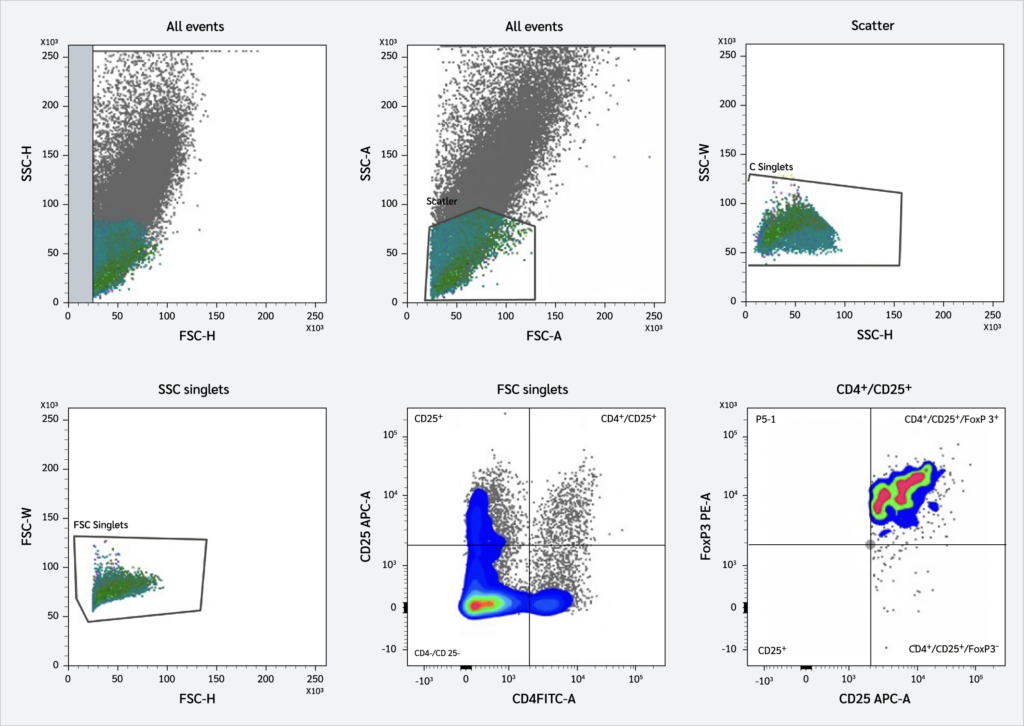 ">
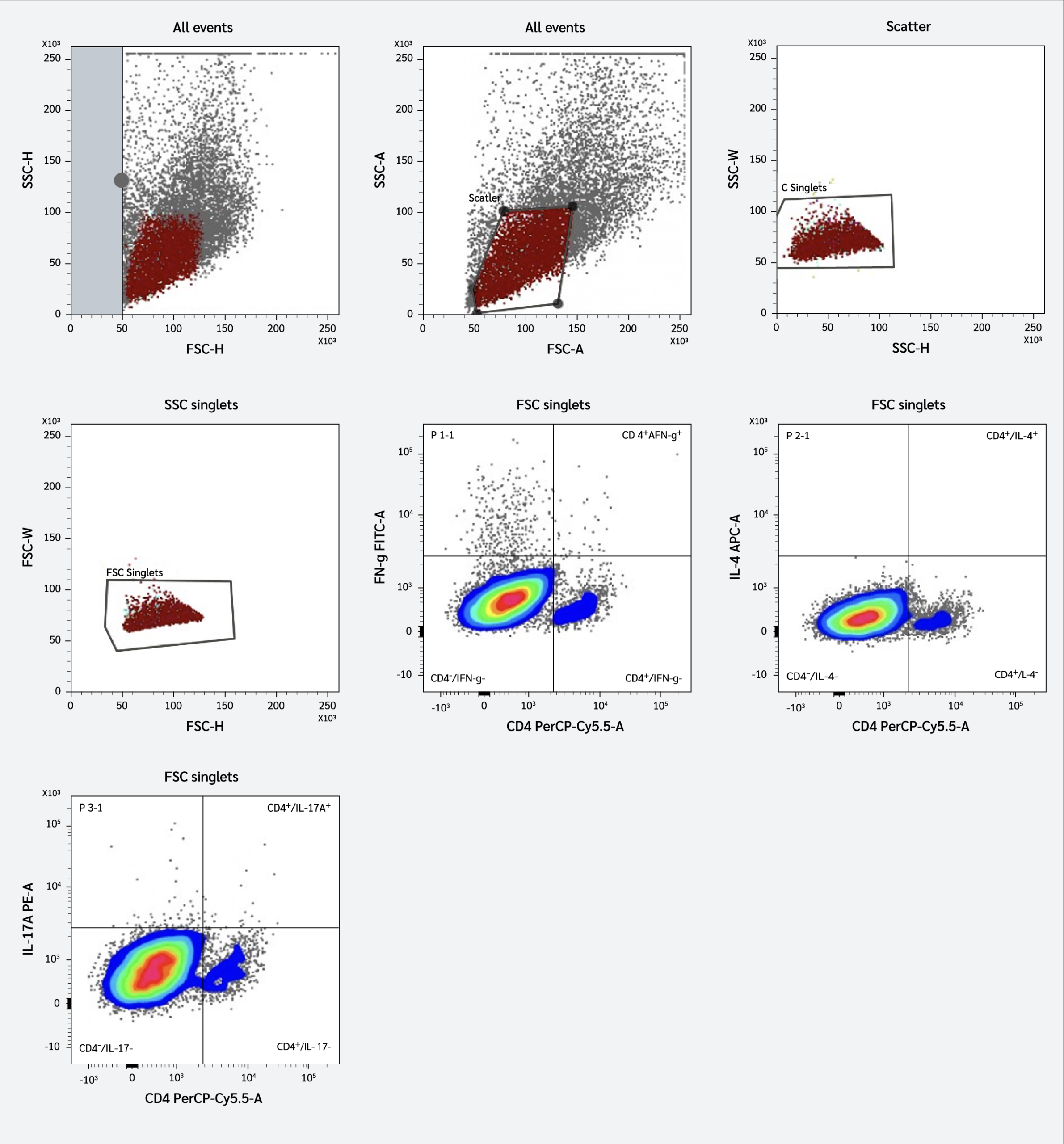
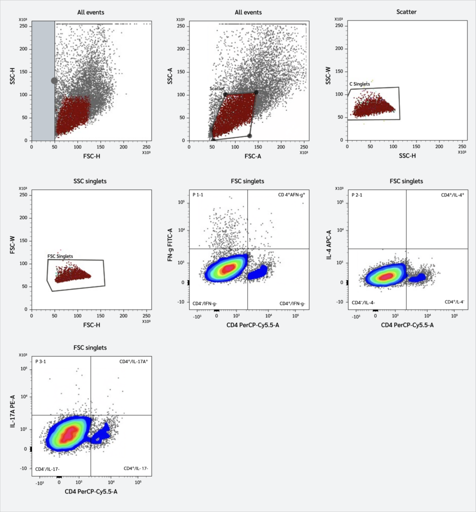
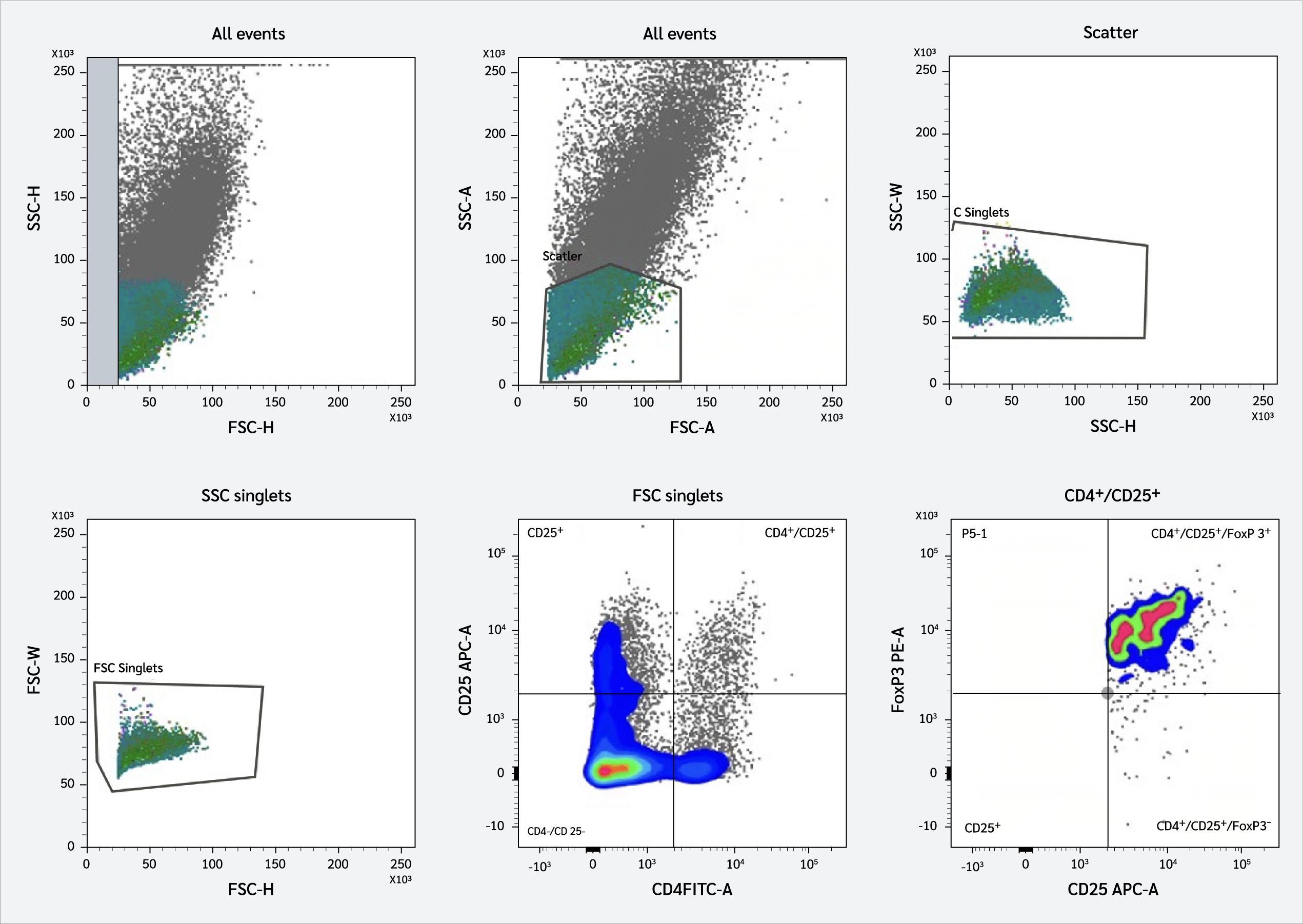
">For Th17 enumeration, the pellet was stained using a Human Th17 Phenotyping Kit (560751; BD Pharmingen, BD Biosciences, San Jose, CA, USA), targeting the CD4+ IL-17A+ population. The detailed hierarchical gating strategy for Th17 cells is shown in Figure 1. For Treg identification, the cells were stained for surface markers CD4 and CD25, followed by intracellular staining for FoxP3 using the Human FoxP3 Buffer Set (560133; BD Pharmingen, BD Biosciences, San Jose, CA, USA), as shown in Figure 2. For both cell populations, fixation and permeabilization were performed using BD Cytofix/Cytoperm™ and Perm/Wash™ buffers to allow antibody access to the intracellular IL-17A and nuclear FoxP3 proteins. Data acquisition was performed on a FACSCalibur flow cytometer and analyzed using CellQuest Pro software, version 5.2.1 (BD Biosciences, San Jose, CA, USA).
Statistical Analysis
All data are presented as mean ± standard deviation (SD). Data distribution was assessed for normality using the Shapiro-Wilk test. Group comparisons were performed using Student’s t test for normally distributed variables. Correlations between variables were analyzed using Spearman’s rank correlation test, and correlation coefficients (r) were reported accordingly. A two-tailed p<0.05 was considered statistically significant. All statistical analyses were conducted using GraphPad Prism software version 10.0.3 (GraphPad Software, San Diego, CA, USA).
Results
Pediatric SLE Patient Characteristics
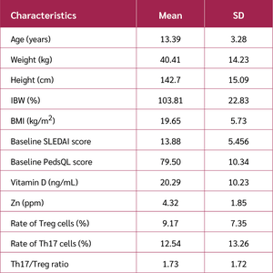
A total of 32 children aged 7.5–18.9 years, with a mean age of 13.39 years, were included in this study. Other patient characteristics are presented in Table 1, while nutritional status is presented in Table 2. Based on the patients’ medical histories, it was noted that none of the recruited subjects received vitamin D supplementation during the study period.
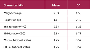
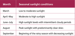
Nutritional status is a complex concept that encompasses a variety of factors in children, including body weight, height, age, and body mass index (BMI). These factors are interrelated and can be used to assess a child’s overall health and well-being and to categorize nutritional status according to the World Health Organization (WHO) and Centers for Disease Control and Prevention (CDC) criteria. Samples were collected from March to September 2025. As a tropical country, Indonesia receives high-intensity sunlight throughout most of the year, particularly between 10:00 AM and 2:00 PM. Detailed information regarding sun exposure is presented in Table 3.
Anthropometric Profile and Growth Distribution
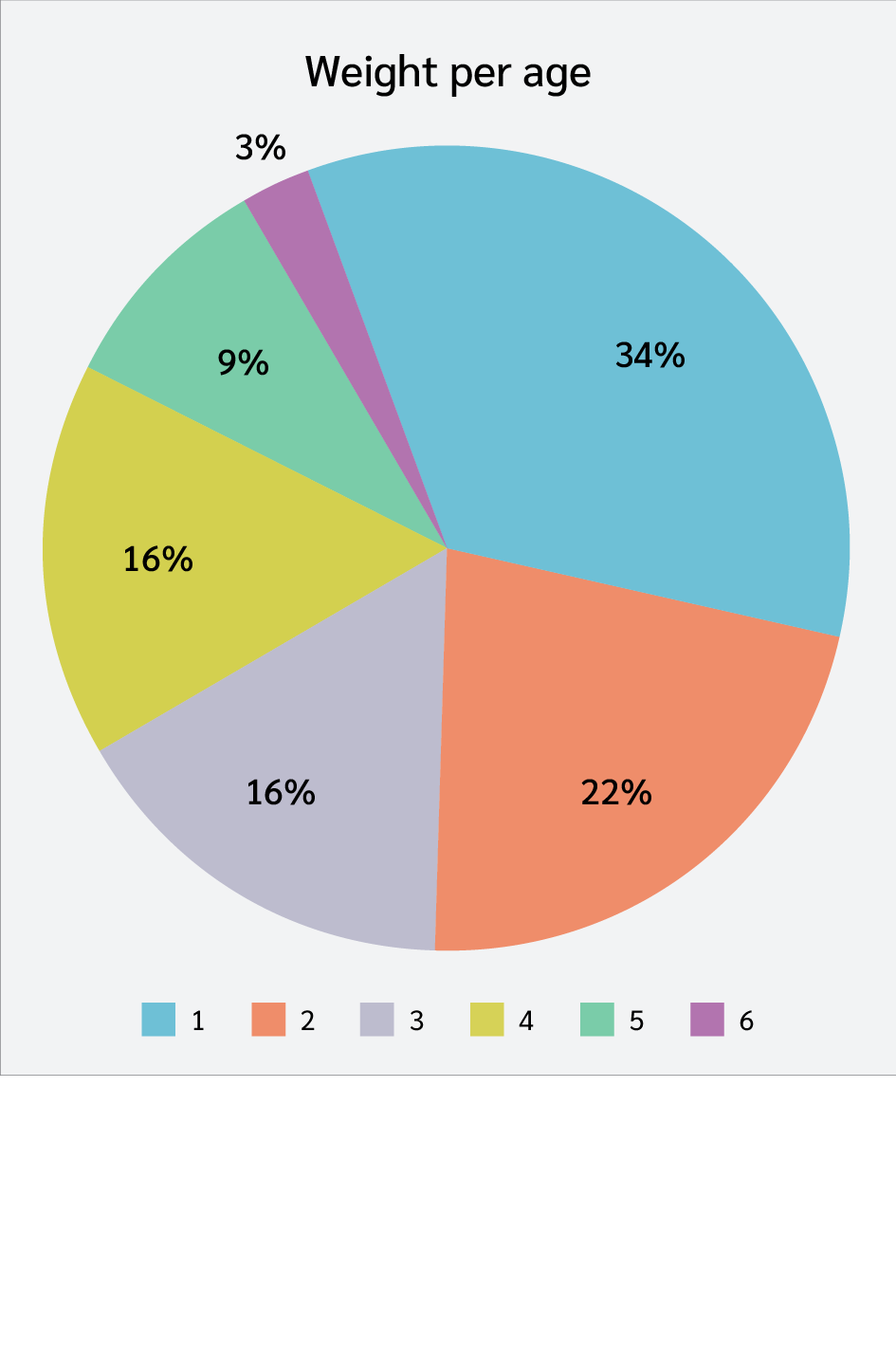
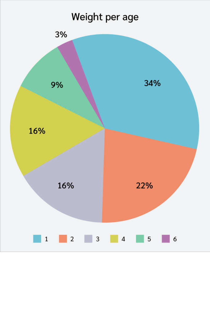
The initial assessment focused on physical development. As shown in Figure 3, the distribution of weight-for-age categories revealed a significant trend toward malnutrition, with the largest proportion of subjects (34%) falling below the 5th percentile (<P5). This was followed by children in the P5–P10 range (22%), while 16% of participants were distributed across each of the P10–P50 categories. In contrast, higher percentiles were less common, with only 12% of the subjects in the P50–P90 range.
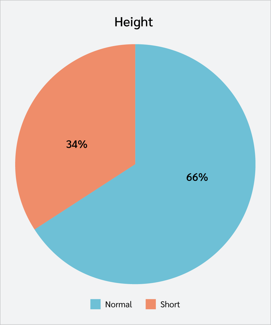
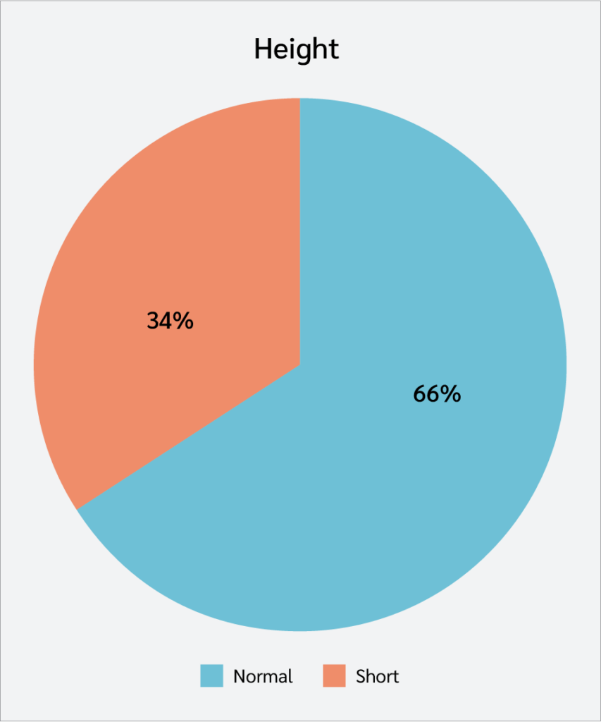
Complementing these findings, Figure 4 presents the distribution of height-for-age. While the majority (66%) exhibited normal height, 34% were classified as having short height. Together, these data indicate a high prevalence of growth impairment and underweight status among the study participants.
Nutritional Status Assessment Based on WHO and CDC Criteria
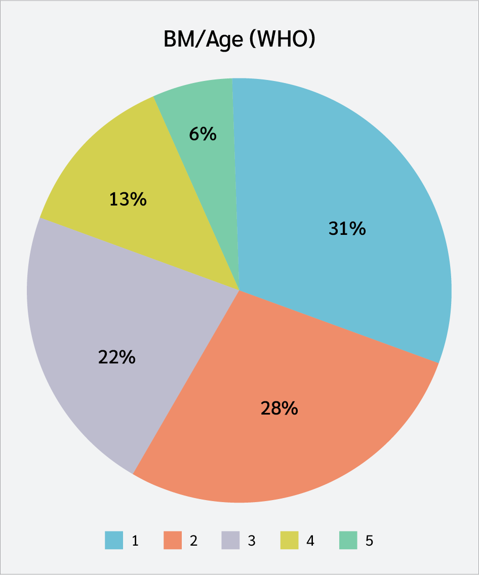
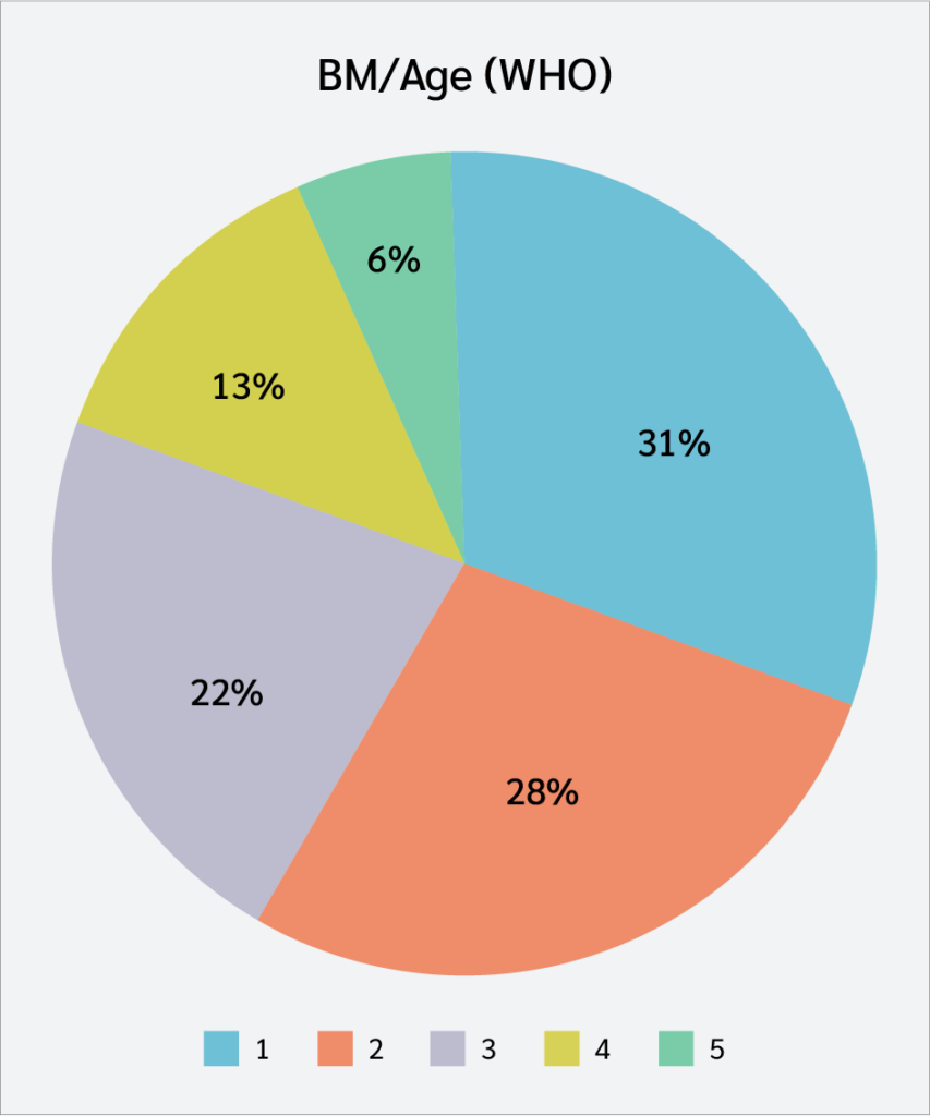
To gain a more detailed understanding of nutritional status, BMI-for-age was further analyzed using both the WHO and CDC growth references. Figure 5 shows the WHO distribution, where the largest group (31%) fell within the −2 to −1 standard deviation (SD) range, indicating that most subjects were toward the lower end of the normal reference range. This pattern remained consistent when applying the CDC growth reference (Figure 6), where the majority of participants also clustered in the lower- to mid-percentile ranges (P5–P75).
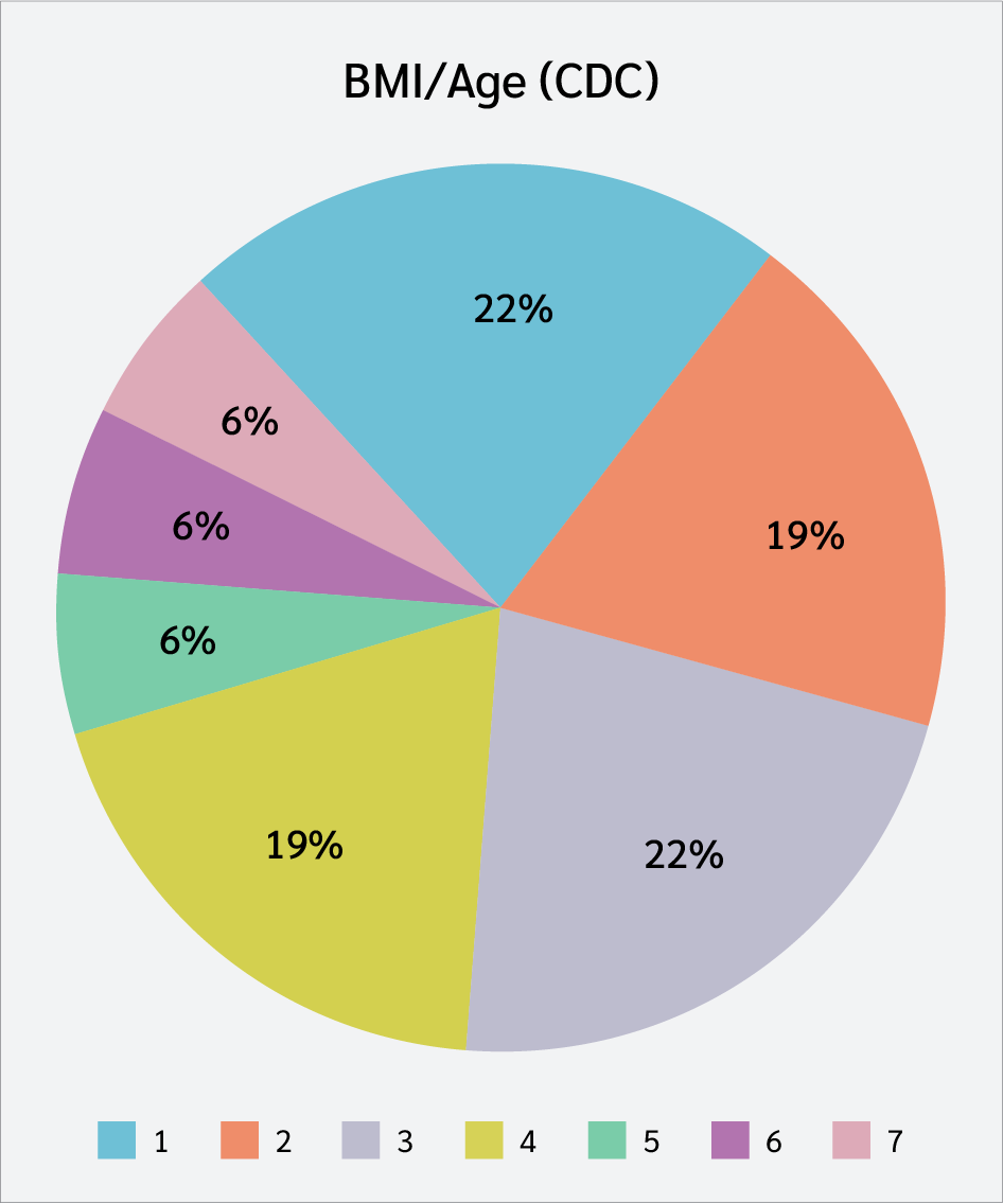
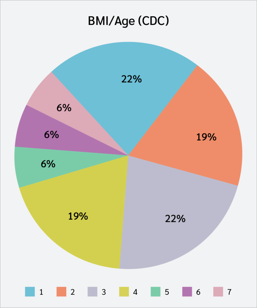
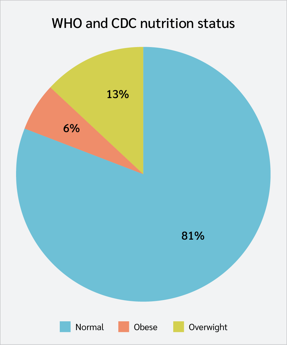
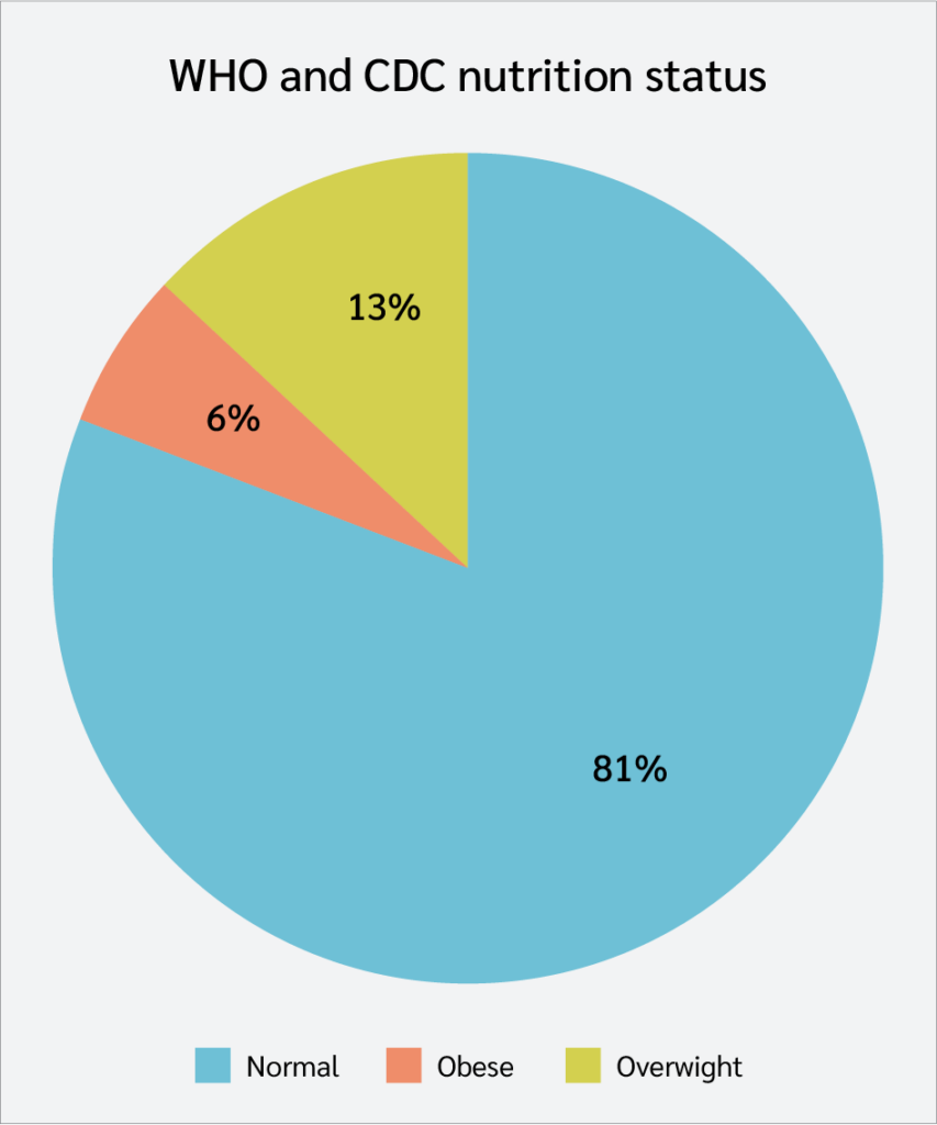
Figure 7 summarizes the overall nutritional status. While 81% of the participants were classified as having normal nutritional status, 19% were categorized as either overweight (13%) or obese (6%). These findings suggest that although underweight-related growth patterns were common, a subset of participants also exhibited excess body weight.
Correlation of SLEDAI Score with Vitamin D and Immunological Parameters
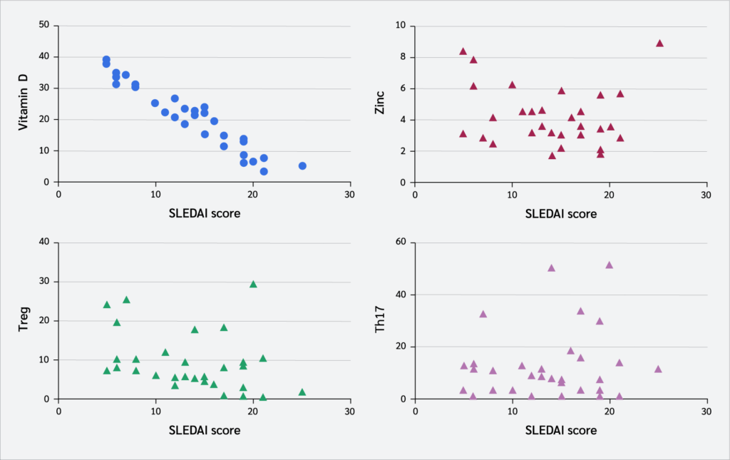 ">
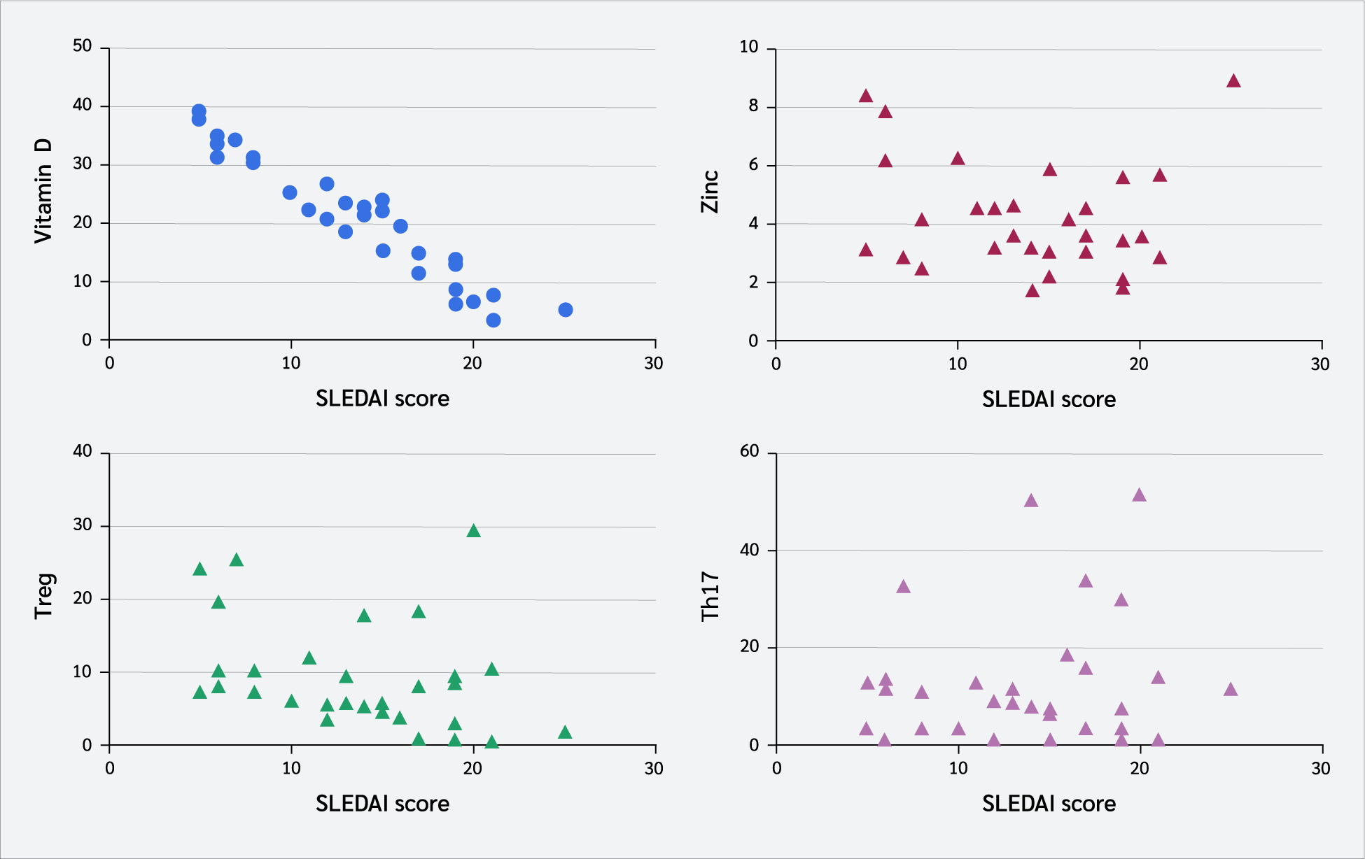
">Beyond physical growth, this study evaluated the relationship between clinical disease activity and biochemical markers. Figure 8 illustrates the relationship between the SLEDAI score and the measured biomarkers, showing a strong inverse correlation with serum vitamin D levels. Unlike zinc levels and the percentages of Treg and Th17 cells, which showed dispersed distributions, vitamin D levels consistently decreased as disease activity increased. Based on serum 25(OH)D levels, the patients were categorized into three groups: 8 (25.0%) with sufficient levels, 9 (28.1%) with insufficient levels, and 15 (46.9%) with deficient levels.
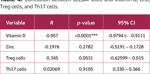
This observation was confirmed by the statistical analysis presented in Table 4. Only vitamin D exhibited a significant relationship with the SLEDAI score (r=-0.957; p<0.0001), with a 95% confidence interval (CI) ranging from -0.9794 – -0.9111. In contrast, no significant correlations were observed between SLEDAI score and zinc levels (r=-0.1976; p=0.2782), Treg cell percentages (r=-0.345; p=0.0531), or Th17 cell percentages (r=0.02069; p=0.9105).
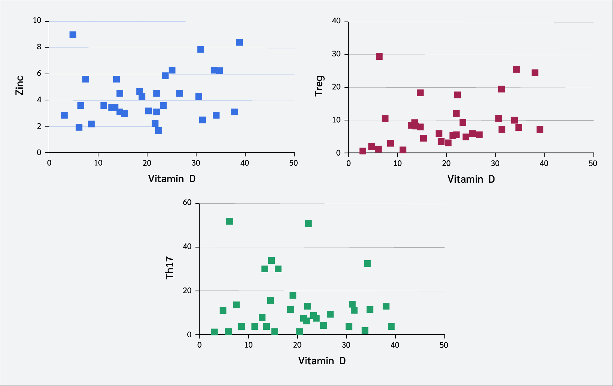
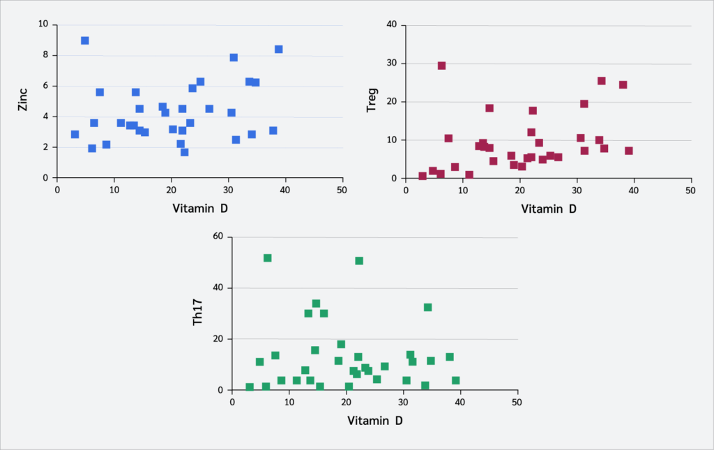
Figure 9 shows the relationship between vitamin D levels and immune cell populations. A positive correlation was observed between vitamin D levels and Treg cell percentages, indicating that higher vitamin D levels were associated with increased Treg cell percentages.
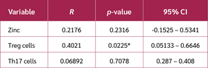
Table 5 confirmed this association. Vitamin D showed a significant positive correlation with Treg cell percentages (r=0.4021; p=0.0225; 95% CI, 0.05133–0.6646), whereas no significant correlations were found with zinc levels (r=0.2176; p=0.2316) or Th17 cell percentages (r=0.06892; p=0.7078). These findings collectively suggest a selective association between vitamin D status and the regulatory immune compartment in patients with SLE.
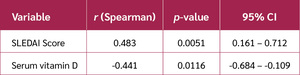 ">
">Consistently, the analysis of the Th17/Treg ratio presented in Table 6 further supported this observation. The Th17/Treg ratio showed a moderate positive correlation with SLEDAI score (r=0.483; p=0.005; 95% CI, 0.161–0.712), indicating that a higher ratio was associated with increased disease activity. In contrast, a moderate negative correlation was observed between the Th17/Treg ratio and vitamin D levels (r=-0.441; p=0.012; 95% CI, -0.684 to -0.109), suggesting that higher vitamin D levels were associated with a lower Th17/Treg ratio.
Discussion
Our findings reveal a significant inverse correlation between serum 25(OH)D concentrations and SLEDAI scores, reinforcing the hypothesis that vitamin D deficiency is associated with increased SLE disease activity (25). In our study, the striking prevalence of suboptimal vitamin D levels, whereby 75% of our pediatric exhibited insufficiency or deficiency, mirrors the high prevalence reported in larger adult cohorts, such as the 170-patient study by Schoindre et al. (26). In the present study, sufficient vitamin D levels (≥30 ng/mL) were identified in only 8 patients (25.0%), whereas vitamin D insufficiency (10≤25(OH)D<30 ng/mL) and deficiency (<10 ng/mL) were found in 9 (28.1%) and 15 patients (46.9%), respectively. The high proportion of patients with suboptimal vitamin D levels further supports the notion that vitamin D depletion is not merely an incidental nutritional deficit but a persistent, disease-related feature of SLE pathophysiology. These findings are reinforced by pediatric-specific data from a cross-sectional study of 45 children with SLE, which demonstrated significantly lower serum 25(OH)D levels than those of 109 healthy controls (14). Similar findings across pediatric and adult populations underscore the high prevalence of vitamin D insufficiency in SLE and suggest that it may reflect both chronic inflammation and disease-related factors.
This observation suggests that reduced vitamin D levels in pediatric SLE cannot be explained solely by environmental or age-related factors. Similar findings have also been reported in Asian populations. A study from Taiwan reported a mean serum vitamin D3 level of 19.7 ± 7.9 ng/mL (27). Collectively, these consistent findings across diverse populations support an association between vitamin D insufficiency and SLE disease activity.
Multiple risk factors predispose patients with SLE to vitamin D deficiency or insufficiency, reflecting the complex interplay between disease characteristics, organ involvement, and therapeutic interventions. One of the principal contributors to vitamin D insufficiency in SLE patients is photosensitivity, a hallmark clinical feature of the disease. Exposure to ultraviolet B (UV-B) radiation can trigger photosensitive reactions, leading patients to avoid sunlight deliberately. In addition to provoking cutaneous manifestations, UV-B exposure has been shown to exacerbate dermatological disorders and precipitate systemic disease flares in SLE (28). Consequently, strict photoprotection practices, although clinically necessary, significantly limit cutaneous vitamin D synthesis and contribute to chronically low serum vitamin D levels.
Beyond photosensitivity, disease-related complications also play an important role in disrupting vitamin D metabolism. Renal involvement, particularly lupus nephritis, is especially important because the kidneys are responsible for the 1α-hydroxylation of vitamin D into its biologically active form. Impaired renal function may therefore reduce the conversion of circulating 25(OH)D to active vitamin D, rendering patients with lupus nephritis especially susceptible to deficiency (29). This mechanism highlights how organ damage intrinsic to SLE can directly influence vitamin D homeostasis.
Pharmacological therapy further compounds this risk. Corticosteroids, which remain a cornerstone of SLE management, adversely affect vitamin D metabolism by reducing intestinal calcium absorption and accelerating the catabolism of both 25(OH)D and 1,25(OH)₂D₃ (30). As a result, patients receiving long-term corticosteroid therapy often exhibit persistently low vitamin D levels despite standard supplementation. Indeed, these patients frequently require higher doses of vitamin D to achieve and maintain adequate serum concentrations (31). Collectively, these findings indicate that vitamin D deficiency in SLE is multifactorial, reflecting the combined effects of disease activity, organ involvement, and treatment-related factors.
Systemic lupus erythematosus (SLE) is characterized by profound immune dysregulation, including the presence of pathogenic autoantibodies, impaired Treg cell function, and expansion of pro-inflammatory Th17 cells. In this context, adequate vitamin D status may help maintain immune homeostasis. Vitamin D participates in antimicrobial defense, macrophage differentiation, and modulation of adaptive immunity, particularly by promoting CD4+ T-cell differentiation toward anti-inflammatory phenotypes such as Th2 and Treg cells. These immunomodulatory properties are essential for preserving immune tolerance and limiting excessive inflammatory responses.
Conversely, vitamin D deficiency may disrupt these regulatory pathways and contribute to immune imbalance. Vitamin D insufficiency has been associated with altered dendritic cell maturation and dysregulated cytokine production, including reduced regulation of key inflammatory cytokines such as IL-6, IL-12, and TNF-α, which are central to SLE pathogenesis (32). Through these mechanisms, low vitamin D levels may facilitate persistent immune activation and autoantibody production.
Clinical interventional studies provide further support for the immunoregulatory role of vitamin D in SLE. A prospective study in patients with hypovitaminosis D receiving 100,000 IU of cholecalciferol weekly for six months demonstrated a significant increase in mean serum 25(OH)D levels, from 18.7 ± 6.7 ng/mL at baseline to 41.5 ± 10.1 ng/mL after treatment (p<0.001) (33). Importantly, vitamin D supplementation was associated with favorable immunological changes, including increased proportions of Treg cells, enhanced FoxP3 and TGFβ expression, expansion of naïve CD4+ T cells, and reductions in memory B cells, Th1 and Th17 effector cells, IL-6 expression, and anti-DNA antibodies (34). Collectively, these findings suggest that vitamin D supplementation may help restore immune homeostasis in patients with SLE.
Evidence regarding the clinical benefits of vitamin D supplementation in SLE continues to evolve, with several studies suggesting a favorable impact on disease activity. One study demonstrated that vitamin D₃ supplementation at a dose of 8000 IU/day for eight weeks in deficient patients and for four weeks in insufficient patients resulted in significant improvement in SLEDAI-2K scores (p=0.028). In the same cohort, anti-double stranded DNA (anti-dsDNA) antibody titers also decreased significantly (p=0.045), suggesting potential immunological benefits alongside clinical improvement (35).
A systematic review further supports these observations. Three out of four included studies reported improvements in SLEDAI scores together with favorable changes in inflammatory markers, fatigue, and endothelial function following vitamin D supplementation. However, one study did not demonstrate a significant improvement in disease activity (36). Moreover, other investigations have failed to identify significant differences between vitamin D supplementation and placebo groups (37). Collectively, these mixed findings indicate that the clinical benefit of vitamin D supplementation may depend on baseline vitamin D status, supplementation dose, treatment duration, and disease heterogeneity, underscoring the need for further well-designed randomized controlled trials.
Conclusion
In conclusion, vitamin D deficiency is common in pediatric SLE and is significantly associated with higher disease activity. Although optimizing vitamin D status may contribute to improved immune homeostasis and disease control, further well-designed prospective studies and randomized controlled trials are needed to clarify the therapeutic role of vitamin D supplementation in pediatric SLE.
Ethical Approval
The study was approved by the Health Research Ethics Commission, Faculty of Medicine, Universitas Brawijaya (Approval No. 208/EC/KEPK/07/2021). It was also registered at ClinicalTrials.gov (Identifier: NCT07069348).
Informed Consent
Written informed consent was obtained from all parents or legal guardians, and informed assent was obtained from all participating children before study enrollment.
Peer-review
Externally peer-reviewed
Author Contributions
Concept – W.B., D.W.; Design – W.B., D.W.; Supervision – W.B.; Funding – W.B., D.W.; Materials – W.B., D.W.; Data Collection and/or Processing – C.M., T.R., R.P.F.; Analysis and/or Interpretation – C.M., T.R.; Literature Review – R.P.F., D.W., C.M., T.R.; Writing – R.P.F., D.W.; Critical Review – W.B.
Conflict of Interest
The authors declared no conflict of interest.
Financial Disclosure:
The authors declared that this study has received no financial support.
AI Statement
The authors used the artificial intelligence (AI) language model Gemini for technical writing assistance. All AI-generated content was critically reviewed and edited by the authors to ensure the accuracy, integrity, and reliability of the manuscript. The authors take full responsibility for the content of the manuscript, and all final decisions, interpretations, and conclusions were made solely by the authors.
Acknowledgment
The authors gratefully acknowledge all the patients and their families for consenting to their children's participation in this study. The authors also thank the medical staff of the Department of Pediatrics at Saiful Anwar Hospital, Malang, and the Biochemical Laboratory, Faculty of Medicine, Universitas Brawijaya, for their invaluable support throughout this study. The contributions of all individuals who provided language editing and proofreading assistance during manuscript preparation are also gratefully acknowledged.
References
Cutolo M, Paolino S, Sulli A, Smith V, Pizzorni C, Seriolo B. Vitamin D, steroid hormones, and autoimmunity. Ann N Y Acad Sci. 2014;1317:39–46. [CrossRef]
Patwardhan VG, Mughal ZM, Padidela R, Chiplonkar SA, Khadilkar VV, Khadilkar AV. Randomized control trial assessing impact of increased sunlight exposure versus vitamin D supplementation on lipid profile in Indian vitamin D deficient men. Indian J Endocrinol Metab. 2017;21(3):393–8. [CrossRef]
Benedik E. Sources of vitamin D for humans. Int J Vitam Nutr Res. 2022;92(2):118–25. [CrossRef]
Weydert JA. Vitamin D in children's health. Children (Basel). 2014;1(2):208–26. [CrossRef]
Aribi M, Mennechet FJD, Touil-Boukoffa C. Editorial: The role of vitamin D as an immunomodulator. Front Immunol. 2023;14:1186635. [CrossRef]
Guritno T, Barlianto W, Wulandari D, Amru WA. Effect of Nigella sativa extract for balancing immune response in pristane induced lupus mice model. J Appl Pharm Sci. 2021;11(7):146–52. [CrossRef]
Levy DM, Kamphuis S. Systemic lupus erythematosus in children and adolescents. Pediatr Clin North Am. 2012;59(2):345–64. [CrossRef]
Tang SP, Lim SC, Arkachaisri T. Childhood-onset systemic lupus erythematosus: Southeast Asian perspectives. J Clin Med. 2021;10(4):559. [CrossRef]
Shamim R, Farman S, Batool S, Khan SEA, Raja MKH. Association of systemic lupus erythematosus disease activity index score with clinical and laboratory parameters in pediatric onset systemic lupus erythematosus. Pak J Med Sci. 2020;36(3):467–72. [CrossRef]
Harry O, Yasin S, Brunner H. Childhood-onset systemic lupus erythematosus: a review and update. J Pediatr. 2018;196:22–30.e2. [CrossRef]
Lai NS, Koo M, Yu CL, Lu MC. Immunopathogenesis of systemic lupus erythematosus and rheumatoid arthritis: the role of aberrant expression of non-coding RNAs in T cells. Clin Exp Immunol. 2017;187(3):327–36. [CrossRef]
Maher SE, Taha HA, Hozayn WG, Okasha AM, Ahmed AE, Shata MA, et al. Study of balance between T-helper 17/T-regulatory cells in systemic lupus erythematosus and its relation to disease activity. Curr Pediatr Res. 2019;23(2):49–55. [CrossRef]
Colaris MJL, van der Hulst RR, Tervaert JWC. Vitamin D deficiency as a risk factor for the development of autoantibodies in patients with ASIA and silicone breast implants: a cohort study and review of the literature. Clin Rheumatol. 2017;36(5):981–93. [CrossRef]
Stagi S, Rigante D. Vitamin D and juvenile systemic lupus erythematosus: Lights, shadows and still unresolved issues. Autoimmun Rev. 2018;17(3):290–300. [CrossRef]
Barragan M, Good M, Kolls JK. Regulation of dendritic cell function by vitamin D. Nutrients. 2015;7(9):8127–51. [CrossRef]
Jiménez-Cortegana C, Palomares F, Alba G, Santa-María C, de la Cruz-Merino L, Sánchez-Margalet V, et al. Dendritic cells: the yin and yang in disease progression. Front Immunol. 2024;14:1321051. [CrossRef]
Iruretagoyena M, Hirigoyen D, Naves R, Burgos PI. Immune response modulation by vitamin D: role in systemic lupus erythematosus. Front Immunol. 2015;6:513. [CrossRef]
Pedro EM, da Rosa Franchi Santos LF, Scavuzzi BM, Iriyoda TMV, Peixe TS, Lozovoy MAB, et al. Trace elements associated with systemic lupus erythematosus and insulin resistance. Biol Trace Elem Res. 2019;191(1):34–44. [CrossRef]
Olza J, Aranceta-Bartrina J, González-Gross M, Ortega RM, Serra-Majem L, Varela-Moreiras G, et al. Reported dietary intake and food sources of zinc, selenium, and vitamins A, E and C in the Spanish population: findings from the ANIBES study. Nutrients. 2017;9(7):697. [CrossRef]
Constantin MM, Nita IE, Olteanu R, Constantin T, Bucur S, Matei C, et al. Significance and impact of dietary factors on systemic lupus erythematosus pathogenesis. Exp Ther Med. 2019;17(2):1085–90. [CrossRef]
Acevedo-Murillo JA, García León ML, Firo-Reyes V, Santiago-Cordova JL, Gonzalez-Rodriguez AP, Wong-Chew RM. Zinc supplementation promotes a Th1 response and improves clinical symptoms in fewer hours in children with pneumonia younger than 5 years old. A randomized controlled clinical trial. Front Pediatr. 2019;7:431. [CrossRef]
Fanouriakis A, Kostopoulou M, Alunno A, Aringer M, Bajema I, Boletis JN, et al. 2019 update of the EULAR recommendations for the management of systemic lupus erythematosus. Ann Rheum Dis. 2019;78(6):736–45. [CrossRef]
Cossarizza A, Chang HD, Radbruch A, Abrignani S, Addo R, Akdis M, et al. Guidelines for the use of flow cytometry and cell sorting in immunological studies (Third edition). Eur J Immunol. 2021;51(12):2708–3145. [CrossRef]
Kim J, Moreno A, Krueger JG. The imbalance between Type 17 T-cells and regulatory immune cell subsets in psoriasis vulgaris. Front Immunol. 2022;13:1005115. [CrossRef]
El-Sherbiny DA, El-Badawy MA, Elmahdi AR. Body mass index in systemic lupus erythematosus: relation to disease activity, bone mineral density and vitamin D level. Egypt J Hosp Med. 2021;82(1):89–95. [CrossRef]
Schoindre Y, Jallouli M, Tanguy ML, Ghillani P, Galicier L, Aumaître O, et al; Group PLUS. Lower vitamin D levels are associated with higher systemic lupus erythematosus activity, but not predictive of disease flare-up. Lupus Sci Med. 2014;1(1):e000027. [CrossRef]
Cheng KH, Tsai MC, Fu LS. The correlation between VitD3 levels and the disease activity of childhood-onset systemic lupus erythematosus. J Chin Med Assoc. 2022;85(5):627–32. [CrossRef]
Justiz Vaillant AA, Goyal A, Varacallo MA. Systemic lupus erythematosus. In: StatPearls [Internet]. Treasure Island (FL): StatPearls Publishing; 2025. [cited January 24, 2026]. Available from: http://www.ncbi.nlm.nih.gov/books/NBK535405/
Li X, Liu J, Zhao Y, Xu N, Lv E, Ci C, et al. 1,25-dihydroxyvitamin D3 ameliorates lupus nephritis through inhibiting the NF-κB and MAPK signalling pathways in MRL/lpr mice. BMC Nephrol. 2022;23(1):243. [CrossRef]
Nowacka A, Śniegocki M, Bożiłow D, Ziółkowska EA. CYP24A1 in small intestinal vitamin D metabolism and clinical implications. Nutrients. 2025;17(21):3348. [CrossRef]
Pludowski P, Wójcik M, Jaworski M, Ochocińska A, Grant WB, Holick MF. Least significant change (LSC) for serum concentrations of 25-hydroxyvitamin D. Nutrients. 2025;17(13):2246. [CrossRef]
Bui L, Zhu Z, Hawkins S, Cortez-Resendiz A, Bellon A. Vitamin D regulation of the immune system and its implications for COVID-19: A mini review. SAGE Open Med. 2021;9:20503121211014073. [CrossRef]
Terrier B, Derian N, Schoindre Y, Chaara W, Geri G, Zahr N, et al. Restoration of regulatory and effector T cell balance and B cell homeostasis in systemic lupus erythematosus patients through vitamin D supplementation. Arthritis Res Ther. 2012;14(5):R221. [CrossRef]
Ghoryani M, Sahebari M, Mahmoudi M, Abdollahi N, Reihani H, Rabe SZT, et al. Immunomodulatory vitamin D effects on regulatory T-cells and cytokines in an in vitro study on patients with systemic lupus erythematosus. Food Agric Immunol. 2016;27(3):377–87. [CrossRef]
Magro R, Saliba C, Camilleri L, Scerri C, Borg AA. Vitamin D supplementation in systemic lupus erythematosus: relationship to disease activity, fatigue and the interferon signature gene expression. BMC Rheumatol. 2021;5(1):53. [CrossRef]
Sousa JR, Rosa ÉPC, Nunes IFOC, Carvalho CMRG. Effect of vitamin D supplementation on patients with systemic lupus erythematosus: a systematic review. Rev Bras Reumatol Engl Ed. 2017;57(5):466–71. English, Portuguese. [CrossRef]
Zheng R, Gonzalez A, Yue J, Wu X, Qiu M, Gui L, et al. Efficacy and safety of vitamin D supplementation in patients with systemic lupus erythematosus: A meta-analysis of randomized controlled trials. Am J Med Sci. 2019;358(2):104–14. [CrossRef]
VOLUME
,
ISSUE
Correspondence
Received
Accepted
Published
Suggested Citation
DOI
License

