
Immune Dysregulation: Mechanisms of Tolerance Failure, Clinical Spectrum, and Emerging Therapeutic Strategies


Abstract
The immune system sustains health through a carefully calibrated equilibrium between activation and restraint, a balance that, when disrupted, gives rise to a clinically heterogeneous spectrum of disorders collectively termed immune dysregulation. Spanning autoimmune diseases, primary and secondary immunodeficiencies, hypersensitivity reactions, and chronic inflammatory states, these conditions collectively affect hundreds of millions of people globally and impose an enormous burden on healthcare systems as well as individual quality of life. Central to the pathophysiology of nearly all immunological disorders is the failure of immune tolerance, including both the thymic mechanisms that eliminate autoreactive lymphocytes during development and the peripheral checkpoints, including regulatory T cells (Tregs), co-inhibitory molecules, and cytokine circuits, that restrain self-reactive clones escaping central deletion. Beyond tolerance failure, immune dysregulation emerges through a convergence of genetic mutations affecting key regulatory genes (e.g., FOXP3, AIRE, RAG, and IL10), epigenetic reprogramming driven by environmental exposures, microbial dysbiosis, and perturbations in cytokine networks. The clinical consequences range from organ-specific destruction in type 1 diabetes mellitus and multiple sclerosis (MS) to life-threatening systemic syndromes including cytokine storm and multi-organ failure. Current therapeutic strategies, including immunomodulatory drugs, biological agents, Janus kinase (JAK) inhibitors, hematopoietic stem cell transplantation (HSCT), and emerging chimeric antigen receptor T (CAR-T)-cell approaches, have transformed outcomes but fall short of providing a cure for most conditions. This review synthesizes the current mechanistic understanding, maps the clinical landscape of immune dysregulation, critically evaluates the evidence for existing and novel therapies, and identifies key gaps where future research must focus to achieve disease-modifying and curative outcomes.
Keywords:
Immune, dysregulation autoimmune, disease immune, tolerance immunodeficiency hypersensitivity cytokine, stormHighlights
Central and peripheral tolerance failure, driven by genetic, epigenetic, and environmental factors, constitutes the common mechanistic thread across the spectrum of immunological disorders.
Loss of FOXP3+ regulatory T-cell function, whether genetic (immune dysregulation, polyendocrinopathy, enteropathy, X-linked [IPEX] syndrome) or acquired, is sufficient to precipitate severe, multisystem autoimmunity, underscoring the indispensable role of active immune suppression.
Cytokine imbalance, characterized by excess tumor necrosis factor-α (TNF-α), interleukin-6 (IL-6), interleukin-17 (IL-17), and interferon-γ (IFN-γ), together with deficient interleukin-10 (IL-10) and transforming growth factor-β (TGF-β), drives chronic inflammation and can escalate to cytokine storm with multi-organ failure.
Janus kinase (JAK) inhibitors, biologic agents targeting specific cytokines or co-stimulatory pathways, and clustered regularly interspaced short palindromic repeats (CRISPR)-based gene correction represent the most promising near-term therapeutic advances.
The role of gut microbial dysbiosis in shaping immune homeostasis remains an underexplored but rapidly evolving area with significant translational potential.
Introduction
Immunology occupies a unique position among the biomedical sciences: it is simultaneously the study of protection and the study of destruction. The very mechanisms that defend the host against an almost infinite variety of microbial threats -pathogen recognition, lymphocyte activation, antibody production, inflammatory cascades- can, under circumstances that are still incompletely understood, turn against the self and cause disease (1,2). The immune system consists of interconnected networks of molecules, proteins, cells, tissues, and organs that work in concert to maintain host integrity against both external pathogens and internal threats (3). This duality is not a design failure; it is an inherent consequence of a system built for exquisite discrimination, where errors at any level exact a biological cost. Immunological disorders, broadly defined as conditions arising from aberrant immune function, encompass autoimmune diseases, primary and secondary immunodeficiencies, hypersensitivity reactions, and chronic inflammatory states (4,5). Their collective global prevalence, which has risen markedly over recent decades, places them among the leading causes of chronic morbidity (6,7). Yet, the mechanistic heterogeneity across and within these categories has historically made unified frameworks difficult to construct.
What unites this clinical diversity at a biological level is the concept of immune homeostasis, the dynamic equilibrium between immune activation and restraint maintained by an intricate architecture of cellular, molecular, and epigenetic regulatory mechanisms (8). When homeostasis breaks down, the pathological consequences reflect the specific layer at which regulation fails: defective thymic selection allows autoreactive lymphocytes to mature; regulatory T-cell (Treg) dysfunction removes the brake on self-reactive responses; cytokine imbalances amplify inflammatory signals beyond the capacity for resolution; and genetic mutations or environmental insults disrupt signaling pathways that normally coordinate immune activity (9-11). The downstream effects are clinically heterogeneous, ranging from the focal destruction of pancreatic β-cells in type 1 diabetes mellitus to the multi-system inflammation of systemic lupus erythematosus (SLE), and from the immunological silence of severe combined immunodeficiency (SCID) to the hyperinflammatory crescendo of cytokine storm syndrome (12-14). Central to virtually all of these conditions is a failure, partial or complete, transient or permanent, of immune tolerance, the state in which the immune system withholds a destructive response against specific antigens (15,16).
Immune tolerance is established and maintained through two complementary layers. Central tolerance, operating in the thymus for T cells and the bone marrow for B cells, eliminates lymphocytes bearing high-affinity receptors for self-antigens through a process of clonal deletion governed in part by the autoimmune regulator (AIRE) gene (17). Peripheral tolerance acts as a secondary checkpoint, encompassing anergy induction, Treg-mediated suppression, and co-inhibitory receptor signaling via cytotoxic T-lymphocyte-associated protein 4 (CTLA-4) and programmed cell death protein 1 (PD-1) (15,18). Regulatory T cells, defined by expression of the master transcription factor FOXP3, are indispensable for active immune suppression. Loss-of-function mutations in FOXP3 cause immune dysregulation, polyendocrinopathy, enteropathy, X-linked (IPEX) syndrome, a devastating neonatal multiorgan autoimmune disease that illustrates the non-redundant requirement for active immune restraint (19,20). Immune dysregulation arises when tolerance mechanisms are overwhelmed, bypassed, or genetically disrupted, resulting in inappropriate immune activation, deficient host defense, or both.
The classification of immunological disorders reflects the predominant mode of immune failure. Hypersensitivity reactions, classified by Gell and Coombs into four types according to their effector mechanisms, represent exaggerated or misdirected responses to antigens that pose no genuine threat (21). Type I (immunoglobulin E [IgE]-mediated) reactions, including asthma, allergic rhinitis, and anaphylaxis, have increased substantially in prevalence over the past half-century, with food allergies disproportionately affecting children (21,22). Autoimmune diseases, affecting an estimated 4–5% of the global population across more than 80 distinct clinical entities, arise from failure of self-tolerance, resulting in immune attack on host tissues (23,24). They display a consistent female predominance, reflecting incompletely understood contributions of sex hormones and X-chromosome-linked immune regulatory genes (25). Immunodeficiency states, whether primary (inborn errors of immunity [IEI]) or secondary (human immunodeficiency virus infection/acquired immunodeficiency syndrome [HIV/AIDS], malnutrition, or iatrogenic), result from inadequate immune responses that permit opportunistic pathogens to establish life-threatening infections (13,26,27). Chronic inflammatory diseases, in which unresolved immune activation damages tissues without a specific self-antigen target, complete this spectrum (28).
Understanding immune dysregulation has accelerated dramatically over the past two decades. The characterization of rare monogenic immunodeficiencies has illuminated critical regulatory nodes (9,29); genome-wide association studies have identified hundreds of susceptibility loci for complex autoimmune diseases (30); and advances in single-cell transcriptomics, epigenomics, and spatial proteomics have revealed cellular heterogeneity at unprecedented resolution. Concurrently, the therapeutic landscape has been transformed by targeted biological agents, small-molecule Janus kinase (JAK) inhibitors, and early-phase immune resetting strategies including chimeric antigen receptor T (CAR-T)-cell therapy (9,31). Yet translating mechanistic depth into clinical benefit remains an ongoing challenge: biologics targeting tumor necrosis factor-α (TNF-α), interleukin-6 (IL-6), and interleukin-17 (IL-17) manage rather than cure immune-mediated diseases; hematopoietic stem cell transplantation (HSCT) offers immune reconstitution with substantial procedural risk; and the prospect of durable remission without continuous immunosuppression, while increasingly plausible, has not yet been achieved at scale (32,33).
This review integrates current mechanistic understanding with the clinical spectrum of immune dysregulation and critically evaluates the evidence base for existing and emerging therapeutic strategies. It identifies the knowledge gaps that constrain progress, particularly the incomplete understanding of tolerance induction in adult tissues, the therapeutic exploitation of the gut microbiome, and the challenge of achieving durable immune reset without global immunosuppression and frames the questions that will define the next decade of immunological investigation.
Immune Tolerance: The Foundation of Self-Discrimination
Central Tolerance
The thymus and bone marrow serve as the primary censors of the immune repertoire, subjecting developing lymphocytes to a dual-selection process that is remarkable in its stringency and, when it fails, in its consequences. In the thymus, as illustrated in Figure 1, developing T cells first undergo positive selection. Only those cells capable of recognizing peptide-major histocompatibility complex (MHC) complexes with sufficient affinity to receive survival signals are permitted to mature, ensuring a self-MHC-restricted repertoire (34). The subsequent step, negative selection, addresses the inherent self-reactivity enriched by positive selection: T cells bearing T-cell receptors (TCRs) with excessively high affinity for self-antigen-MHC complexes are eliminated by apoptosis (clonal deletion), a process mediated in part by medullary thymic epithelial cells and dendritic cells (3).
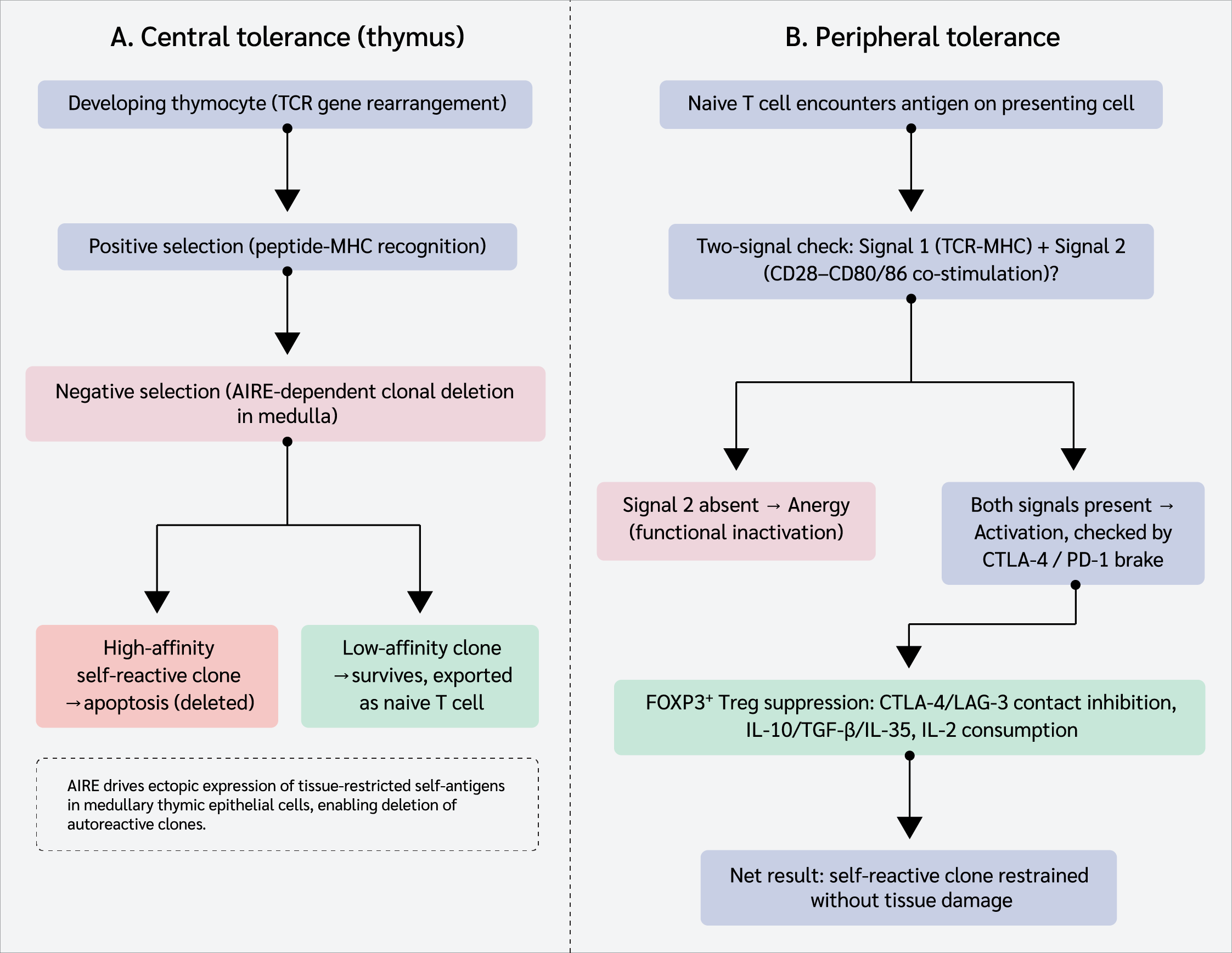
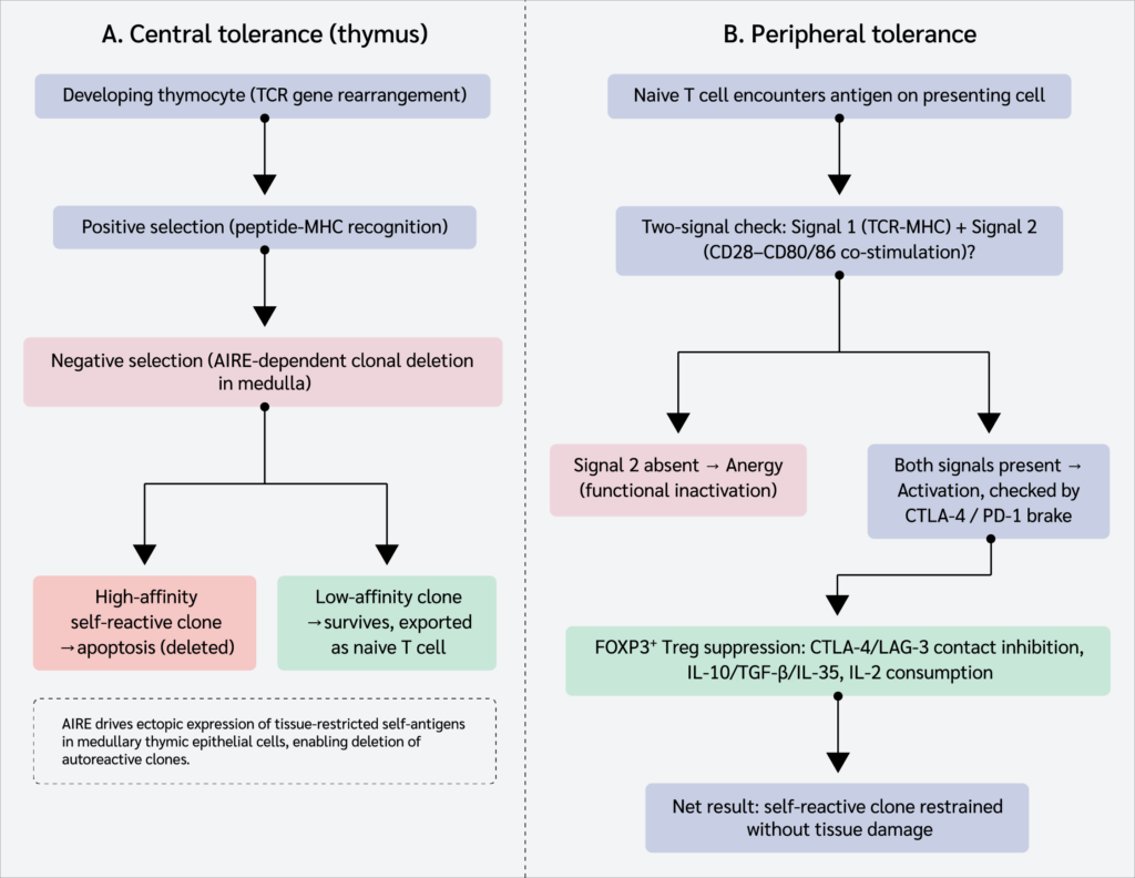
Central to thymic negative selection is the AIRE gene, whose product drives the ectopic expression of thousands of tissue-restricted antigens within medullary thymic epithelial cells. Mutations in AIRE cause autoimmune polyendocrinopathy-candidiasis-ectodermal dystrophy (APECED), illustrating with near-experimental precision the indispensability of this mechanism for self-tolerance. Analogous central processes operate in the bone marrow for B lymphocytes: autoreactive B cells undergo receptor editing or clonal deletion, and those that survive carry receptors of sufficiently low self-antigen affinity that peripheral regulatory mechanisms can normally contain them (15,34).
Peripheral Tolerance
No central selection process operates with perfect efficiency, and autoreactive lymphocytes circulate in healthy individuals without causing disease, a testament to the robustness of peripheral tolerance. The principal peripheral mechanisms are anergy (functional inactivation through the absence of co-stimulation), clonal deletion, and active suppression by FOXP3+ Tregs (15,18). The two-signal model of lymphocyte activation is fundamental here: Signal 1 (MHC-TCR binding) in the absence of Signal 2 (cluster of differentiation 80 [CD80]/CD86-CD28 co-stimulation) induces anergy rather than activation. Negative regulators, most notably CTLA-4 on Tregs, which competes with CD28 for CD80/CD86 with far higher affinity, function as physiological brakes on T-cell activation (15).
These FOXP3+ Tregs deploy multiple suppressive mechanisms: contact inhibition via CTLA-4 and lymphocyte activation gene 3 (LAG-3), immunosuppressive cytokines (IL-10, TGF-β, and IL-35), and competitive consumption of IL-2 (18,20). The centrality of FOXP3 to immune homeostasis is illustrated by IPEX syndrome, caused by loss-of-function mutations in FOXP3, which produces severe neonatal diabetes, inflammatory enteropathy, eczema, and multi-organ autoimmunity, often fatal in infancy without HSCT (19,20). Additionally, IL-10-producing type 1 regulatory T (Tr1) cells and TGF-β-producing T helper 3 (Th3) cells represent distinct regulatory lineages that complement canonical FOXP3+ Treg function (18). Immune homeostasis dysregulation arising from these failures can lead to a range of pathologies from debilitating autoimmune diseases to severe immunodeficiencies (8).
Mechanisms of Immune Dysregulation
Genetic Architecture of Immune Dysregulation
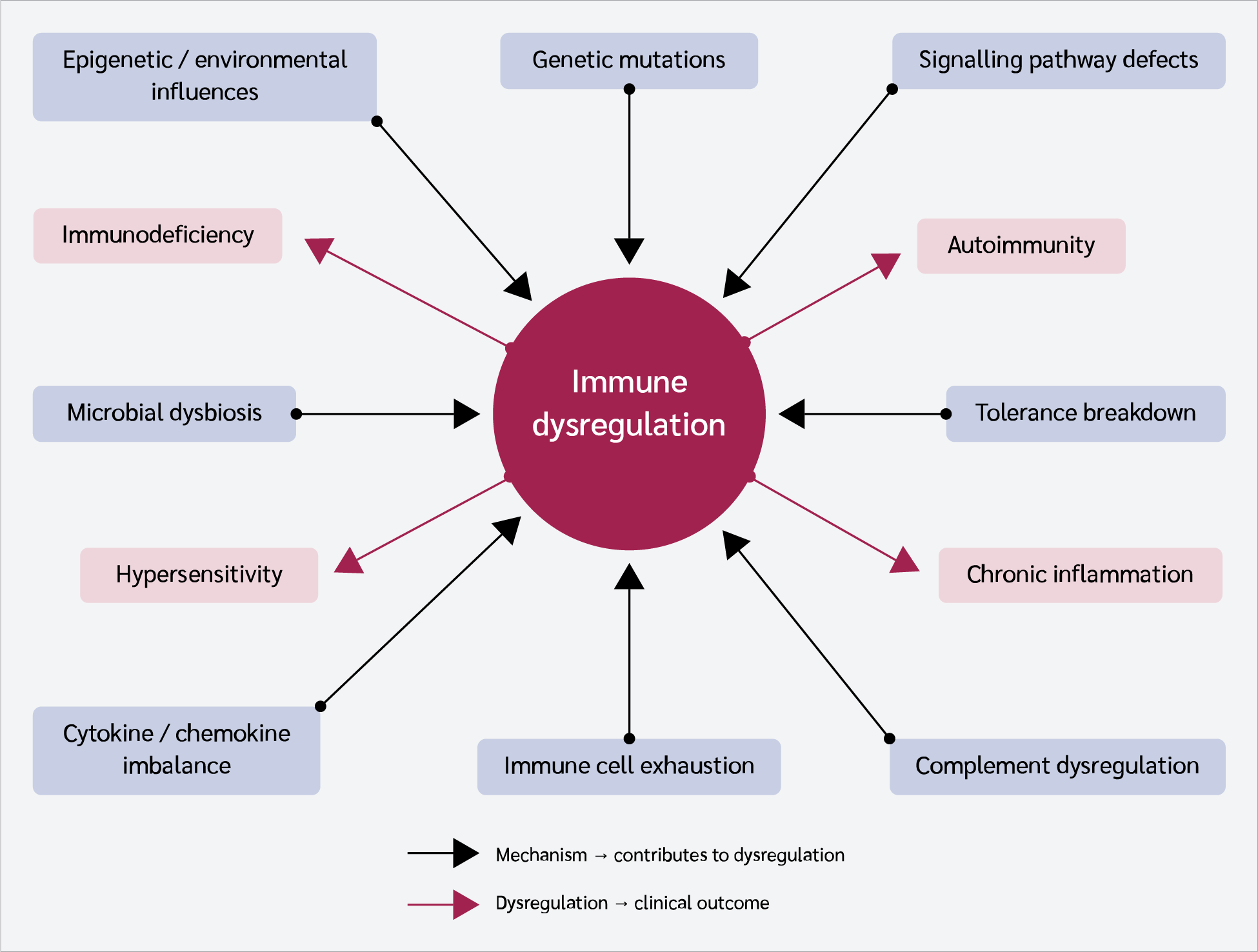
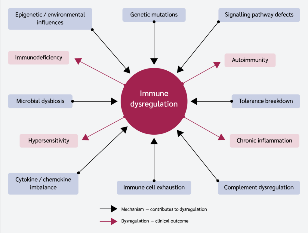
Single-gene disorders have delineated the non-redundant functions of immune regulatory molecules with remarkable clarity. Severe combined immunodeficiency (SCID), caused by mutations in genes encoding components of the TCR signaling pathway or the common γ-chain of cytokine receptors, ablates T-cell development and leaves affected infants profoundly susceptible to life-threatening infections (35,36). Immune dysregulation, polyendocrinopathy, enteropathy, X-linked (IPEX) syndrome arises from mutations in the FOXP3 gene, as described in Figure 2. Type I hypersensitivity susceptibility has been associated with gain-of-function mutations in caspase recruitment domain family member 11 (CARD11) and loss-of-function mutations in mucosa-associated lymphoid tissue lymphoma translocation protein 1 (MALT1), both converging on aberrant nuclear factor kappa B (NF-κB) signaling in lymphocytes (37,38). Mutations in recombination-activating genes (RAG) have been associated with autoimmunity (9), while damaging mutations in interleukin-10 (IL10), interleukin-10 receptor subunit alpha (IL10RA), and interleukin-10 receptor subunit beta (IL10RB) cause severe, early-onset inflammatory bowel disease (IBD) (39-41). Genome-wide association studies of SLE have identified hundreds of susceptibility loci, many in genes encoding complement components and interferon-pathway regulators (5,30). While mutations in these genes or pathways predispose to immune dysregulation, environmental triggers are essential for progression to clinical disease (9,11).
Epigenetic and Environmental Contributions
Epigenetic modifications, including DNA methylation, histone acetylation, microRNA dysregulation, and chromatin remodeling, alter gene expression in immune cells without changing the underlying DNA sequence, and accumulating evidence implicates them in immune dysregulation (6,42). In SLE, hypomethylation of interferon-stimulated genes in T cells has been documented, and in vitro demethylation has been shown to induce autoantibody production, a causal link between epigenetic state and autoimmune potential (42). Environmental exposures serve as epigenetic triggers in genetically predisposed individuals: ultraviolet B radiation activates keratinocyte apoptosis and nucleosomal antigen release in SLE, as shown in Figure 2; Epstein-Barr virus (EBV) molecular mimicry between EBV nuclear antigen 1 (EBNA-1) and the Ro/La autoantigens represents one of the strongest epidemiological links between infection and systemic autoimmunity; and chemicals such as hydralazine and procainamide induce drug-related lupus through demethylation of immune regulatory genes (5,30). Dietary patterns exert immunomodulatory effects both directly and indirectly through the gut microbiota and its metabolites, with high-fat, low-fiber diets reducing circulating short-chain fatty acids (SCFAs) that normally drive Treg differentiation (10).
Microbial Dysbiosis and Mucosal Immunity
The gut harbors the largest concentration of immune cells in the body, and the continuous dialogue between commensal microbiota and intestinal immune cells shapes systemic immune tone throughout life (43). Commensal microbes drive intestinal Treg differentiation, mucosal tolerance, and appropriate activation thresholds; dysbiosis, defined as qualitative and quantitative shifts in microbial community composition, has been associated with IBD, rheumatoid arthritis (RA), multiple sclerosis (MS), and SLE (43). Mechanistically, altered microbial metabolites, such as SCFAs, can disrupt epithelial integrity and immune signaling pathways. Persistent infectious agents, most notably Mycobacterium tuberculosis and HIV, additionally cause T-cell exhaustion characterized by progressive loss of cytokine secretion and sustained upregulation of PD-1 and CTLA-4, which compromises both pathogen clearance and immune regulation (27,44). Molecular mimicry between microbial antigens and host proteins represents another pathway to tolerance breakdown, generating cross-reactive immune responses that inadvertently target self-tissues following infection (5).
Cytokine and Chemokine Imbalance
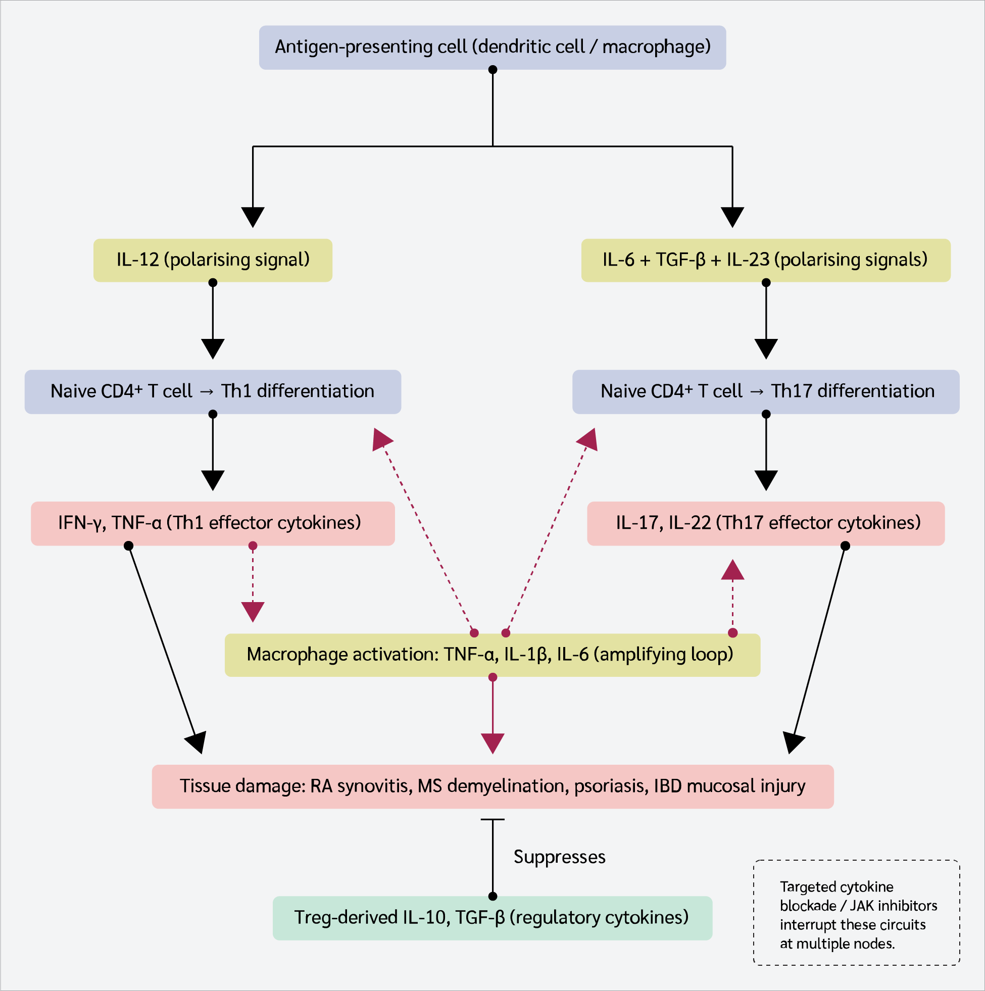
Cytokines are not merely biomarkers of immune activity; they are the causal agents of the tissue damage and systemic symptoms that define immune-mediated diseases (9,21). The pathogenic cytokine landscape in autoimmunity is dominated by TNF-α, IL-1β, IL-6, IL-17, and IFN-γ, which collectively promote dendritic cell maturation, Th17 polarization, macrophage activation, and osteoclast activation, as shown in Figure 3. Dysregulation of type I interferons and pro-inflammatory cytokines, particularly IL-6 and TNF-α, plays a critical role in SLE pathogenesis (30,45). Dysregulation of IL-1 production causes autoinflammatory diseases including deficiency of IL-1 receptor antagonist and cryopyrin-associated periodic syndromes (CAPS) (21,29). Disruption of key signaling pathways such as NF-κB and JAK-signal transducer and activator of transcription (STAT) leads to abnormal immune activation or suppression (9). In extreme cases, dysregulated cytokine release culminates in a cytokine storm, a hyperinflammatory state characterized by systemic inflammation, multiorgan dysfunction, and high mortality (12,46).
Immune Cell Dysfunction and the Exhaustion Paradigm
Effective immunity depends not merely on the magnitude of the immune response but on the functional integrity of its cellular constituents. T-cell exhaustion, arising from chronic antigen stimulation, is defined by graded loss of effector functions, reduced proliferative capacity, and increased expression of inhibitory receptors including T-cell immunoglobulin and mucin-domain containing-3 (TIM-3), T-cell immunoreceptor with immunoglobulin and immunoreceptor tyrosine-based inhibitory motif domains (TIGIT), PD-1, LAG-3, and CTLA-4 (44). B-cell dysregulation contributes to autoimmunity through loss of tolerance checkpoints that allow autoreactive germinal center reactions, producing high-affinity autoantibodies that mediate pathology through fragment crystallizable (Fc) receptor activation and complement fixation (23). In SLE, dysfunction of B cells, T cells, and dendritic cells, together with impaired clearance of apoptotic cells, collectively amplifies inflammation (30,45). Dysfunction of B-cell and T-cell subsets, particularly Tregs and Th17 cells, as well as dendritic cells, contributes to the inflammatory milieu characteristic of SLE and other systemic autoimmune diseases (5).
Complement System Dysregulation
The complement system, activated through the classical, lectin, and alternative pathways, serves as a crucial effector arm of innate immunity facilitating pathogen clearance, opsonization, and inflammation (2,21). Deficiencies in early complement components (C1q, C2, and C4) are paradoxically associated with SLE through impaired immune complex clearance, while uncontrolled activation, as in atypical hemolytic uremic syndrome caused by factor H mutations, drives endothelial injury through deposition of the membrane attack complex (2). Regulatory proteins including factor H and decay-accelerating factor (DAF) are essential for preventing excessive complement activation on host tissues; mutations or functional impairments in these regulators tip the balance toward pathological inflammation. These mechanisms of immune dysregulation rarely operate in isolation; their convergence in genetically susceptible individuals exposed to environmental triggers exemplifies the multifactorial complexity of immunological disorders, underscoring the need for systems-level approaches to research and management (9,11).
Classification and Clinical Spectrum of Immunological Disorders
Hypersensitivity Reactions: Exaggerated Immune Activation
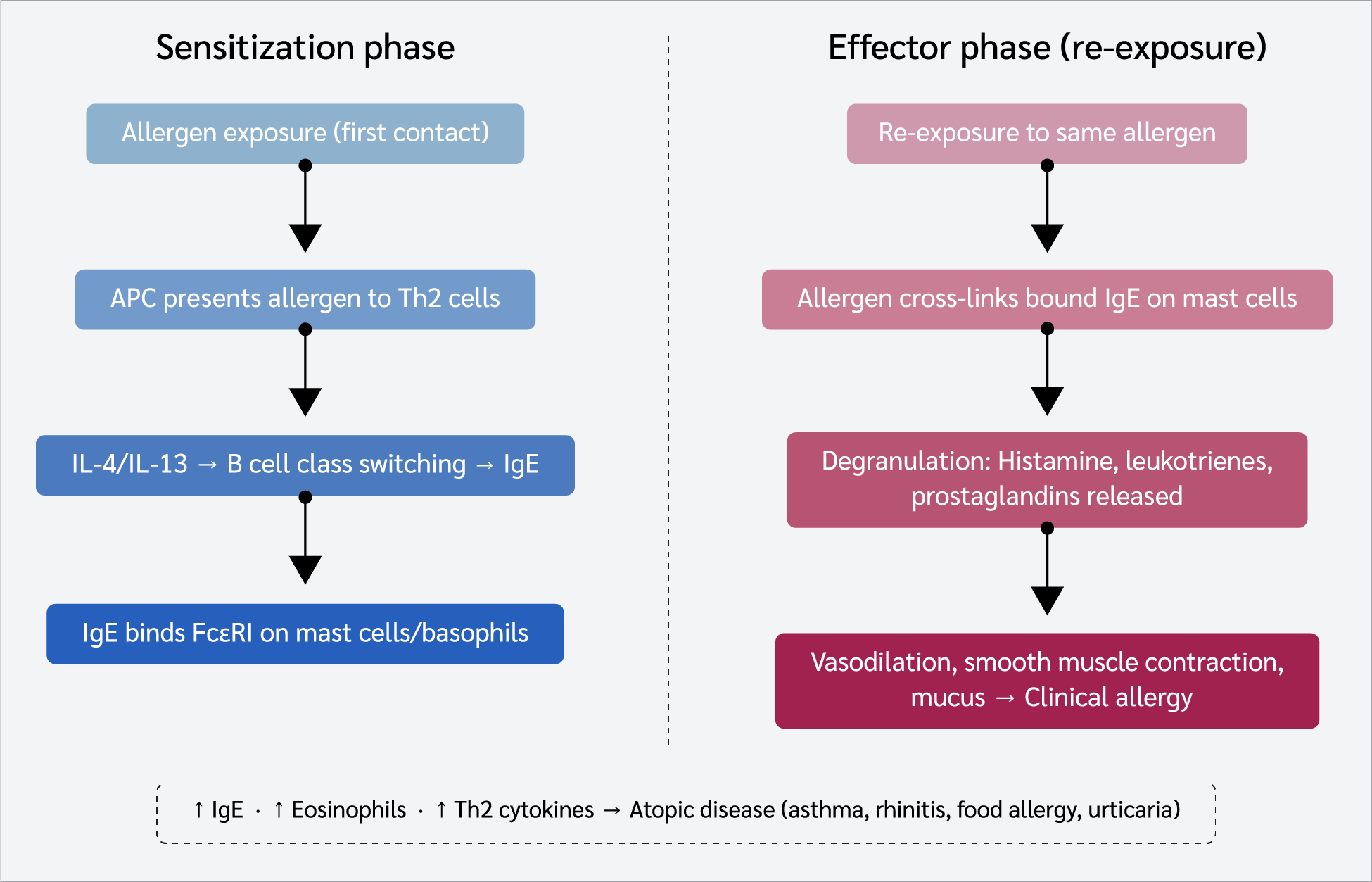
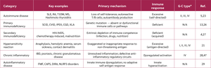
The Gell and Coombs classification (summarized in Table 1) retains conceptual utility as an organizing framework for the diverse mechanisms by which inappropriate immune responses against innocuous antigens cause pathology (21). Type I hypersensitivity reactions, illustrated in Figure 4, are mediated by allergen-specific IgE cross-linking of FcεRI receptors on mast cells and basophils and are the most clinically prevalent. Their global increase has been attributed to the hygiene hypothesis, positing that reduced exposure to commensal organisms deprives the immune system of regulatory signals that normally constrain Th2 responses (22,48). Type II reactions, mediated by cytotoxic IgG or IgM antibodies directed against cell-surface antigens, operate through complement activation and antibody-dependent cellular cytotoxicity (ADCC), exemplified by autoimmune hemolytic anemia, Goodpasture syndrome, myasthenia gravis, and Graves disease (21). Type III (immune complex-mediated) reactions are illustrated by SLE, serum sickness, and reactive arthritis, and are characterized by complement activation and neutrophil recruitment at sites of complex deposition (21). Type IV delayed-type hypersensitivity, driven by antigen-experienced T cells, manifests 48–72 hours after antigen challenge, underpinning contact dermatitis, tuberculin reactions, and graft rejection (3,21).
This framework has been refined over the past decade. The original hygiene hypothesis, framed around reduced childhood infection, has been largely superseded by the biodiversity hypothesis, which attributes the rise in Type I hypersensitivity less to fewer infections per se and more to reduced early-life exposure to the broader range of environmental and commensal microorganisms that historically calibrated regulatory immune development; this distinction matters clinically, since it reframes prevention away from deliberately permitting infection and toward restoring microbial diversity, while also helping explain why some epidemiological data linking infection frequency directly to allergic disease risk have been inconsistent across cohorts.
Autoimmune Diseases: Tolerance Failure at Scale
More than 80 distinct autoimmune entities are recognized, collectively affecting approximately 4–5% of the global population with a consistent female predominance (4,24,25). All share a breakdown in the distinction between self and non-self, though their underlying mechanisms, effector pathways, and target organs are highly heterogeneous (5,23). Autoantibodies function as pharmacological agents at their targets: in Graves disease, thyroid-stimulating hormone (TSH)-receptor antibodies act as agonists driving thyroid overactivity; in myasthenia gravis, acetylcholine receptor antibodies act as antagonists blocking neuromuscular transmission (3). In SLE, nuclear antigen-antibody immune complexes deposit in glomeruli, skin, and synovium, activating complement and recruiting neutrophils that release neutrophil extracellular traps (NETs). These structures expose additional nuclear antigens, creating a self-amplifying autoimmune cycle (5,14). Helper CD4+ T cells, particularly Th17 cells producing IL-17, are key pathogenic effectors in RA, MS, psoriasis, and IBD, while FOXP3+ Treg cells that would normally restrain these responses appear to adopt a dysfunctional pro-inflammatory phenotype, a phenomenon designated Treg plasticity, whose molecular basis remains incompletely understood (4,9).
This literature is not fully settled, however. Human fate-mapping and lineage-tracing studies have produced conflicting estimates of how frequently bona fide FOXP3-stable Tregs convert to an IL-17-producing, pro-inflammatory phenotype in vivo, as opposed to a minority of unstable, low-FOXP3 cells that are selectively expanded under inflammatory conditions. This distinction has materially different therapeutic implications: the former would argue for stabilizing existing Tregs, while the latter would argue for excluding unstable clones from adoptive Treg therapy protocols. Reconciling these positions, rather than treating Treg plasticity as an established fact, remains an open methodological priority for the field.
Immunodeficiency: The Consequences of Immune Absence
Primary immunodeficiencies, now formally designated inborn errors of immunity (IEIs), encompass over 450 recognized disorders affecting innate immunity, adaptive immunity, immune regulation, or combinations thereof (13,26). Their clinical presentations reflect the specific immunological compartment affected: antibody deficiencies (such as X-linked agammaglobulinemia [XLA] and common variable immunodeficiency [CVID]) manifest with recurrent sinopulmonary infections; T-cell and combined immunodeficiencies confer susceptibility to viral, fungal, and opportunistic pathogens; and complement deficiencies predispose to Neisseria infections and autoimmune diseases through impaired immune complex clearance (2,26). Secondary immunodeficiencies, which are more prevalent globally, arise from HIV infection, iatrogenic immunosuppression, and malnutrition, each creating predictable patterns of immune deficiency (4,27). Infection with HIV remains paradigmatic: selective CD4+ T-cell depletion disrupts coordinated adaptive immune responses, permits opportunistic infections, and results in chronic immune activation that accelerates non-infectious morbidities (27).
Clinical Manifestations of Immune Dysregulation
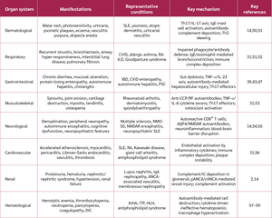
The clinical manifestations of immune dysregulation are shaped by three intersecting variables: the nature of the immune defect (excessive, deficient, or misdirected), the organ systems targeted, and the phase of disease. Organ-specific manifestations serve as important diagnostic indicators of the underlying immunological disorder, as mapped out in Table 2 (2,24,49). Dermatological manifestations frequently serve as the diagnostic entry point, particularly in SLE and psoriasis, where the Th17-mediated keratinocyte hyperproliferation at sites of mechanical trauma (the Koebner phenomenon) provides visible evidence of systemic immune dysregulation (14,50). Neurological involvement has emerged as an especially dynamic field, with the discovery of specific neural autoantibodies transforming the classification and management of autoimmune encephalitis, a condition previously misdiagnosed as psychiatric disorders (14,55). Severe complications, cytokine storm syndrome, hemophagocytic lymphohistiocytosis, and acute respiratory distress syndrome, represent the extreme consequence of immune regulatory failure and are associated with high short-term mortality (12,57,60). In cytokine storm, positive feedback loops between immune cells result in persistent cytokine secretion and overwhelming systemic inflammation, driving vascular leakage, coagulopathy, and multi-organ failure (12,46,61).
Prevention, Treatment, and Emerging Therapeutic Strategies
Therapeutic strategy in immune dysregulation is shaped by two competing imperatives: the need to suppress pathological immune responses and the imperative to preserve host defense against infection and malignancy (31). Most current treatments resolve this tension imperfectly. The clinical success of TNF-α blockade in RA, IBD, and psoriasis provides direct pharmacological proof that cytokine excess drives tissue pathology. Janus kinase inhibitors provide a valuable oral therapeutic option with broader cytokine inhibition but carry class-specific risks, including thromboembolism, which remain under active regulatory scrutiny, as listed in Table 3 (9). Hematopoietic stem cell transplantation remains the only reliably curative approach for many monogenic immunodeficiencies and severe autoimmune phenotypes, but its risk-benefit profile constrains its use to conditions sufficiently severe to justify procedural mortality; low survival and increased rates of graft failure have been reported in some subsets (9,32). Abatacept and belatacept have shown promising results in CTLA-4 deficiency as first-line therapy to control immune dysregulation, though increased infection susceptibility during treatment remains a concern for long-term use (32,33). Gene therapy has achieved regulatory approval for adenosine deaminase (ADA)-SCID and is in advanced trials for recombination-activating gene 1/recombination-activating gene 2 severe combined immunodeficiency (RAG1/RAG2 SCID), chronic granulomatous disease, and Wiskott-Aldrich syndrome; its autologous nature eliminates graft-versus-host disease (9,13). Early-phase trials of anti-CD19 CAR-T-cell therapy in refractory SLE and myositis have generated extraordinary excitement with preliminary durable drug-free remissions, but replication in larger cohorts is needed (9). Immunomodulatory drugs, including ruxolitinib for inflammatory and proliferative disease, novel peptide drugs, and anti-T-cell antibodies, expand the pharmacological toolkit, while prevention strategies, including vaccination, dietary microbiome intervention, and neonatal SCID screening (via T-cell receptor excision circle [TREC] assays) broaden the public health approach to immune dysregulation (9,26).
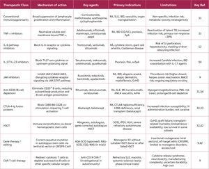
It is worth being explicit about the limits of this evidence base at the present time: the published experience consists of small, uncontrolled, single-arm case series from a handful of centers, typically including fewer than 20 patients per report, with follow-up rarely exceeding two to three years; no randomized controlled trial has yet been reported, the durability of remission beyond this window is unknown, and the risk of long-term B-cell aplasia or secondary malignancy with CAR-T-cell therapy in a nonmalignant, potentially younger population has not been systematically characterized. These findings should accordingly be considered as hypothesis-generating rather than practice-changing evidence at this stage.
This regulatory scrutiny was substantially sharpened by the ORAL Surveillance trial, a large, randomized safety study in RA patients aged 50 years or older with cardiovascular risk factors, which found higher rates of major adverse cardiovascular events, malignancy, and venous thromboembolism with tofacitinib compared with TNF-α inhibitors, prompting boxed-warning label changes for the entire JAK inhibitor class. Whether this risk generalizes to younger, lower-cardiovascular-risk patients, and to other diseases treated with JAK inhibitors beyond RA, remains actively debated, and current guidance favors reserving JAK inhibitors for patients in whom TNF-α inhibitors have failed or are contraindicated, rather than using them as a first-line biologic-sparing option.
Future Directions and Knowledge Gaps
Despite remarkable advances in understanding and treating immune dysregulation, the field faces several fundamental challenges. Single-cell multi-omics technologies, combining transcriptomics, epigenomics, proteomics, and spatial profiling, are beginning to reveal the heterogeneity of immune states across disease stages and between individuals, potentially enabling prediction of disease trajectories before clinical manifestation. The prospect of durable immune reset, rather than ongoing suppression, is supported by CAR-T-cell therapy data. Regulatory T-cell therapeutics, including expanded polyclonal Treg infusions and antigen-specific engineered Tregs, are currently in clinical trials as biologically elegant approaches to restoring immune regulation. The microbiome-immune interface holds substantial promise: fecal microbiota transplantation is well established in IBD, and its application in systemic autoimmunity is under active investigation. Artificial intelligence (AI) and machine learning applied to multi-modal immunological datasets are producing predictive models for disease flares, treatment response, and at-risk-to-disease transition whose clinical implementation will require careful attention to interpretability and regulatory frameworks. With advances in technologies such as spatial transcriptomics and AI, further breakthroughs in immunology are anticipated. Molecular therapies, including monoclonal antibodies, B-cell depleting agents, and CAR-T-cell treatments, represent a step toward more precisely targeted therapies with potentially fewer off-target side effects; however, as detailed below, this potential remains to be confirmed in controlled trials, and a cure for most autoimmune diseases remains an aspiration rather than a demonstrated outcome.
Controversies and Evidence Gaps
Several topics covered in this review remain genuinely unsettled rather than representing incremental refinements of an agreed understanding and are highlighted here explicitly rather than being folded silently into the narrative above. First, whether Treg dysfunction in autoimmune disease principally reflects numerical deficiency, functional impairment of otherwise normal-numbered Tregs, or outright lineage instability (Treg plasticity) remains contested, and different answers imply different therapeutic strategies (Treg expansion, Treg stabilization, or exclusion of unstable clones from adoptive therapy). Second, the JAK inhibitor cardiovascular and malignancy signal identified in ORAL Surveillance was generated in an older, cardiovascular-risk-enriched RA population, and whether it applies with similar magnitude across the younger, more diverse populations now being treated with JAK inhibitors for atopic dermatitis, alopecia areata, and inflammatory bowel disease is an open, clinically consequential question. Third, the CAR-T-cell literature in autoimmunity, discussed above, currently rests entirely on small, uncontrolled case series; the field lacks any randomized comparison against existing biologic therapy, and claims of a durable immune reset should therefore be regarded as preliminary until such data become available. Finally, the therapeutic microbiome literature is asymmetric: fecal microbiota transplantation has strong evidence in Clostridioides difficile infection and some support in ulcerative colitis, but its extension to systemic autoimmune disease rests largely on small pilot studies and preclinical models and should not be presented with the same evidentiary weight as the gut-specific indications. We identify these four areas as priorities for future systematic review and, where feasible, randomized controlled trials, rather than as settled components of current practice.
Conclusion
Immune dysregulation is not a single disease but a mechanistic category encompassing the full spectrum of conditions that arise when the immune system loses its capacity for discriminating, proportionate, and self-limited responses. The shared language of tolerance failure, cytokine dysregulation, cellular exhaustion, and genetic predisposition connects conditions as clinically diverse as IPEX syndrome, SLE, allergic asthma, and HIV-associated immunodeficiency into a coherent framework that illuminates both pathogenesis and therapeutic logic. The past decade has brought transformative advances: biological agents that precisely interrupt individual cytokine pathways, JAK inhibitors that broadly dampen cytokine signaling, and the first glimpses of curative approaches through gene therapy and immune resetting strategies. To date, no curative therapy is available for most autoimmune diseases. However, recent research indicates that molecular therapies, monoclonal antibodies, B-cell depleting agents, and, more speculatively, CAR-T-cell treatments may benefit patients by addressing some limitations of traditional approaches.
Nevertheless, as discussed above, the evidence for several of these newer strategies remains preliminary, and a cure for most autoimmune and immunodeficiency conditions has not yet been demonstrated at scale. The path toward closing persistent gaps depends on mechanistic clarity regarding how genetic susceptibility, environmental exposures, and microbial ecology converge on threshold-dependent transitions from immune health to immune disease, together with therapeutic innovation in antigen-specific tolerance induction, Treg-based therapies, and clustered regularly interspaced short palindromic repeats (CRISPR)-enabled gene correction. Achieving durable, curative outcomes will require the full integration of cellular, molecular, and clinical immunology with the technological and analytical tools that are already reshaping what is possible to know and to do about the human immune system.
Ethical Approval
Not applicable.
Informed Consent
Not applicable.
Peer-review
Externally peer-reviewed
Author Contributions
Concept – A.M., A.S.A., S.H.A., A.A.G., B.R.O., A.R.A.; Design – A.M., A.S.A., S.H.A., A.A.G., B.R.O., A.R.A.; Supervision – A.S.A.; Literature Review – A.M., A.S.A., S.H.A., A.A.G., B.R.O., A.R.A.; Writing – A.S.A., A.M.; Critical Reviews – A.M., A.S.A., S.H.A., A.A.G., B.R.O., A.R.A.
Conflict of Interest
The authors declared that they have no conflict of interest.
Financial Disclosure:
The authors declared that this study has received no financial support.
AI Statement
During the preparation of this manuscript, the authors used Claude AI to obtain additional information and Microsoft Copilot to generate images. The authors reviewed and edited the generated content as necessary and take full responsibility for the content of this publication.
References
Misekova G, Geldiyeva G, Annamyradova M. Immunology: a comprehensive overview of the immune system and its functions. Obrazovanie i Nauka v XXI Veke. 2025;59–4(1).
Murphy K, Weaver C. Janeway's immunobiology. 10th ed. New York (NY): Garland Science; 2022.
Muñoz-Carrillo JL, Castro-García FP, Chávez-Rubalcaba F, Chávez-Rubalcaba I, Martínez-Rodríguez JL, Hernández-Ruiz ME. Immune system disorders: hypersensitivity and autoimmunity. In: Athari SS, editor. Immunoregulatory Aspects of Immunotherapy [Internet]. London: IntechOpen; 2018 [cited June 26, 2026]. Available from: https://www.intechopen.com/chapters/60540
Starshinova AA, Savchenko AA, Borisov A, Kudryavtsev I, Rubinstein A, Dovgalyuk I, et al. Immunological disorders: gradations and the current approach in laboratory diagnostics. Pathophysiology. 2025;32(2):17. [CrossRef]
Albarbar B, Aga H. A review on autoimmune diseases: recent advances and future perspectives. AlQalam J Med Appl Sci. 2024;7(3):718–29. [CrossRef]
Nautiyal G, Sharma I, Pandey P, Sharma SK. Autoimmune diseases: recent insights on epidemiology, pathogenesis, and prevalence rate. In: Raza K, Singh S, eds. Artificial Intelligence and Autoimmune Diseases. Singapore: Springer, 2024: 33–58.
Borisov AG. Clinical characterization of functional disorders affecting immune system. Med Immunol (Russ). 2013;15(1):45–50. [CrossRef]
Wang L, Liang Y, Zhao C, Ma P, Zeng S, Ju D, et al. Regulatory T cells in homeostasis and disease: molecular mechanisms and therapeutic potential. Signal Transduct Target Ther. 2025;10(1):345. [CrossRef]
Taietti I, Catamerò F, Lodi L, Giovannini M, Castagnoli R. Inborn errors of immunity with atopic phenotypes in the allergy and immunology clinic: a practical review. Curr Opin Allergy Clin Immunol. 2025;25(2):105–14. [CrossRef]
Arneth B. Molecular Mechanisms of Immune Regulation: A Review. Cells. 2025;14(4):283. [CrossRef]
Gambineri E, Torgerson TR. Genetic disorders with immune dysregulation. Cell Mol Life Sci. 2012;69(1):49–58. [CrossRef]
Fajgenbaum DC, June CH. Cytokine Storm. N Engl J Med. 2020;383(23):2255–73. [CrossRef]
Notarangelo LD, Bacchetta R, Casanova JL, Su HC. Human inborn errors of immunity: an expanding universe. Sci Immunol. 2020;5(49):eabb1662. [CrossRef]
Tsokos GC. Autoimmunity and organ damage in systemic lupus erythematosus. Nat Immunol. 2020;21(6):605–14. [CrossRef]
Meng X, Layhadi JA, Keane ST, Cartwright NJK, Durham SR, Shamji MH. Immunological mechanisms of tolerance: Central, peripheral and the role of T and B cells. Asia Pac Allergy. 2023;13(4):175–86. [CrossRef]
Bluestone JA, Anderson M. Tolerance in the Age of Immunotherapy. N Engl J Med. 2020;383(12):1156–66. [CrossRef]
Anderson MS, Venanzi ES, Klein L, Chen Z, Berzins SP, Turley SJ, et al. Projection of an immunological self shadow within the thymus by the aire protein. Science. 2002;298(5597):1395–401. [CrossRef]
Ferreira LMR, Muller YD, Bluestone JA, Tang Q. Next-generation regulatory T cell therapy. Nat Rev Drug Discov. 2019;18(10):749–69. [CrossRef]
Ochs HD, Gambineri E, Torgerson TR. IPEX, FOXP3 and regulatory T-cells: a model for autoimmunity. Immunol Res. 2007;38(1–3):112–21. [CrossRef]
Rudensky AY. Regulatory T cells and Foxp3. Immunol Rev. 2011;241(1):260–8. [CrossRef]
Marshall JS, Upton JEM, Vliagoftis H, Hildebrand KJ, Byrne A, Watson W. Introduction to immunology and immune disorders. Allergy Asthma Clin Immunol. 2024;20(Suppl 3):69. [CrossRef]
Lyons JJ, Milner JD. Primary atopic disorders. J Exp Med. 2018;215(4):1009–22. [CrossRef]
Sciascia S, Bizzaro N, Meroni PL, Dimitrios B, Borghi MO, Bossuyt X, et al. Autoantibodies testing in autoimmunity: diagnostic, prognostic and classification value. Autoimmun Rev. 2023;22(7):103356. [CrossRef]
Pisetsky DS. Pathogenesis of autoimmune disease. Nat Rev Nephrol. 2023;19(8):509–24. [CrossRef]
Sasikala K, Kumari MR. An account on promising alternative system of medicine to boost immunity. Plant Sci Today. 2022;9(Suppl 2):47–50. [CrossRef]
Tangye SG, Al-Herz W, Bousfiha A, Cunningham-Rundles C, Franco JL, Holland SM, et al. Human inborn errors of immunity: 2022 update on the classification from the International Union of Immunological Societies Expert Committee. J Clin Immunol. 2022;42(7):1473–507. [CrossRef]
Tuano KS, Seth N, Chinen J. Secondary immunodeficiencies: an overview. Ann Allergy Asthma Immunol. 2021;127(6):617–26. [CrossRef]
Medzhitov R. Origin and physiological roles of inflammation. Nature. 2008;454(7203):428–35. [CrossRef]
Delmonte OM, Castagnoli R, Calzoni E, Notarangelo LD. Inborn errors of immunity with immune dysregulation: from bench to bedside. Front Pediatr. 2019;7:353. [CrossRef]
Albarbar B, Aga H, Shalbi SM. Systemic lupus erythematosus: an overview of pathogenesis, prevalence, diagnosis, classification criteria, management, challenges, and future directions. Khalij-Libya J Dent Med Res. 2024;8(2):207–13. [CrossRef]
McInnes IB, Gravallese EM. Immune-mediated inflammatory disease therapeutics: past, present and future. Nat Rev Immunol. 2021;21(10):680–6. [CrossRef]
Slatter MA, Engelhardt KR, Burroughs LM, Arkwright PD, Nademi Z, Skoda-Smith S, et al. Hematopoietic stem cell transplantation for CTLA4 deficiency. J Allergy Clin Immunol. 2016;138(2):615–9.e1. [CrossRef]
Lo B, Zhang K, Lu W, Zheng L, Zhang Q, Kanellopoulou C, et al. Patients with LRBA deficiency show CTLA4 loss and immune dysregulation responsive to abatacept therapy. Science. 2015;349(6246):436–40. [CrossRef]
Hogquist KA, Baldwin TA, Jameson SC. Central tolerance: learning self-control in the thymus. Nat Rev Immunol. 2005;5(10):772–82. [CrossRef]
Bacchelli C, Moretti FA, Carmo M, Adams S, Stanescu HC, Pearce K, et al. Mutations in linker for activation of T cells (LAT) lead to a novel form of severe combined immunodeficiency. J Allergy Clin Immunol. 2017;139(2):634–42.e5. [CrossRef]
Punwani D, Wang H, Chan AY, Cowan MJ, Mallott J, Sunderam U, et al. Combined immunodeficiency due to MALT1 mutations, treated by hematopoietic cell transplantation. J Clin Immunol. 2015;35(2):135–46. [CrossRef]
Dadi H, Jones TA, Merico D, Sharfe N, Ovadia A, Schejter Y, et al. Combined immunodeficiency and atopy caused by a dominant negative mutation in caspase activation and recruitment domain family member 11 (CARD11). J Allergy Clin Immunol. 2018;141(5):1818–30.e2. [CrossRef]
McKinnon ML, Rozmus J, Fung SY, Hirschfeld AF, Del Bel KL, Thomas L, et al. Combined immunodeficiency associated with homozygous MALT1 mutations. J Allergy Clin Immunol. 2014;133(5):1458–62, 1462.e1–7. [CrossRef]
Glocker EO, Kotlarz D, Boztug K, Gertz EM, Schäffer AA, Noyan F, et al. Inflammatory bowel disease and mutations affecting the interleukin-10 receptor. N Engl J Med. 2009;361(21):2033–45. [CrossRef]
Glocker EO, Frede N, Perro M, Sebire N, Elawad M, Shah N, et al. Infant colitis—it's in the genes. Lancet. 2010;376(9748):1272. [CrossRef]
Kotlarz D, Beier R, Murugan D, Diestelhorst J, Jensen O, Boztug K, et al. Loss of interleukin-10 signaling and infantile inflammatory bowel disease: implications for diagnosis and therapy. Gastroenterology. 2012;143(2):347–55. [CrossRef]
Hedrich CM. Mechanistic aspects of epigenetic dysregulation in SLE. Clin Immunol. 2018;196:3–11. [CrossRef]
Belkaid Y, Hand TW. Role of the microbiota in immunity and inflammation. Cell. 2014;157(1):121–41. [CrossRef]
Wherry EJ, Kurachi M. Molecular and cellular insights into T cell exhaustion. Nat Rev Immunol. 2015;15(8):486–99. [CrossRef]
Nagy G, Koncz A, Perl A. T- and B-cell abnormalities in systemic lupus erythematosus. Crit Rev Immunol. 2005;25(2):123–40. [CrossRef]
Shimabukuro-Vornhagen A, Gödel P, Subklewe M, Stemmler HJ, Schlößer HA, Schlaak M, et al. Cytokine release syndrome. J Immunother Cancer. 2018;6(1):56. [CrossRef]
Xavier RJ, Podolsky DK. Unravelling the pathogenesis of inflammatory bowel disease. Nature. 2007;448(7152):427–34. [CrossRef]
Thomsen SF. Epidemiology and natural history of atopic diseases. Eur Clin Respir J. 2015;2:24642. [CrossRef]
Abbas AK, Lichtman AH, Pillai S. Cellular and molecular immunology. 10th ed. Philadelphia (PA): Elsevier; 2021.
Lowes MA, Suárez-Fariñas M, Krueger JG. Immunology of psoriasis. Annu Rev Immunol. 2014;32:227–55. [CrossRef]
Lambrecht BN, Hammad H. The immunology of asthma. Nat Immunol. 2015;16(1):45–56. [CrossRef]
Bonilla FA, Barlan I, Chapel H, Costa-Carvalho BT, Cunningham-Rundles C, de la Morena MT, et al. International Consensus Document (ICON): Common variable immunodeficiency disorders. J Allergy Clin Immunol Pract. 2016;4(1):38–59. [CrossRef]
Smolen JS, Aletaha D, McInnes IB. Rheumatoid arthritis. Lancet. 2016;388(10055):2023–38. Erratum in: Lancet. 2016;388(10055):1984. [CrossRef]
Reich DS, Lucchinetti CF, Calabresi PA. Multiple Sclerosis. N Engl J Med. 2018;378(2):169–80. [CrossRef]
Heneka MT, Carson MJ, El Khoury J, Landreth GE, Brosseron F, Feinstein DL, et al. Neuroinflammation in Alzheimer's disease. Lancet Neurol. 2015;14(4):388–405. [CrossRef]
Libby P, Buring JE, Badimon L, Hansson GK, Deanfield J, Bittencourt MS, et al. Atherosclerosis. Nat Rev Dis Primers. 2019;5(1):56. [CrossRef]
Jordan MB, Allen CE, Weitzman S, Filipovich AH, McClain KL. How I treat hemophagocytic lymphohistiocytosis. Blood. 2011;118(15):4041–52. [CrossRef]
Al-Samkari H, Berliner N. Hemophagocytic lymphohistiocytosis. Annu Rev Pathol. 2018;13:27–49. [CrossRef]
Delves PJ, Martin SJ, Burton DR, Roitt IM. Roitt's essential immunology. 14th ed. Hoboken (NJ): Wiley-Blackwell; 2021.
Thompson BT, Chambers RC, Liu KD. Acute respiratory distress syndrome. N Engl J Med. 2017;377(6):562–72. [CrossRef]
Hotchkiss RS, Monneret G, Payen D. Immunosuppression in sepsis: a novel understanding of the disorder and a new therapeutic approach. Lancet Infect Dis. 2013;13(3):260–8. [CrossRef]
VOLUME
,
ISSUE
Correspondence
Received
Accepted
Published
Suggested Citation
DOI
License

