
Maternal Gut Dysbiosis Exacerbates Offspring Susceptibility to Collagen-Induced Arthritis in Mice


Abstract
Objective:
The gut microbiota plays a critical role in immune system development and the maintenance of immune tolerance. Dysbiosis, particularly during the maternal period, has been implicated in autoimmune diseases. This study investigated whether maternal antibiotic-induced dysbiosis exacerbates collagen-induced arthritis (CIA) in offspring and whether fecal microbiota transplantation (FMT) can reverse this effect.
Materials and Methods:
To establish a dysbiosis model, pregnant BALB/c mice were administered a combination of amoxicillin (40 mg/kg) and vancomycin (30 mg/kg) from the last week of pregnancy until the male pups used in the experiment (n=5 per group) reached 8 weeks of age (total treatment duration, 9 weeks). In the FMT group, oral FMT was administered after completion of antibiotic therapy. The bacterial profiles of the mice gut microbiota were analyzed by sequencing, and CIA was subsequently induced in all groups.
Results:
Maternal dysbiosis significantly reduced bacterial populations in the gut microbiota, whereas FMT restored these populations. Additionally, dysbiosis was associated with increased arthritis severity in the CIA model, whereas FMT appeared to reduce it. These results demonstrate that the maternal gut microbiota plays a critical role in the pathogenesis of autoimmune arthritis and that FMT may represent a potential therapeutic approach.
Conclusion:
Maternal dysbiosis in the gut microbiota and its treatment with FMT affected body weight in mice, particularly causing low birth weight. Dysbiosis increased disease severity in the CIA model, whereas FMT treatment reduced it.
Keywords:
Gut, microbiota dysbiosis fecal, microbiota, transplantation collagen-induced, arthritis rheumatoid, arthritis autoimmunityIntroduction
The critical role of the gut microbiota in organismal homeostasis and immune system development is increasingly being understood through numerous studies (1,2). This microbial diversity plays a central role in the development of immune tolerance, in addition to performing essential functions such as nutrient metabolism, colonization resistance against pathogens, and immunomodulation (3,4). Dysbiosis, which refers to a disruption in the functional balance of the gut microbiota, is associated with various inflammatory and autoimmune diseases (5).
Factors in early life are particularly important in the development of dysbiosis. Colonization of the microbiota begins in the prenatal period, and maternal factors such as the mother's diet, medications (particularly antibiotics), and delivery mode shape the newborn's microbial composition (6,7). Studies have shown that dysbiosis caused by maternal antibiotic use disrupts intestinal barrier integrity, thereby triggering systemic inflammation and having long-term effects on immunological maturation (8,9). This is particularly important in the pathogenesis of autoimmune diseases, which are characterized by impaired immune tolerance.
Rheumatoid arthritis (RA), a chronic, systemic autoimmune disease, can develop as a result of dysbiosis (10,11). In addition to genetic predisposition, environmental triggers play a role in the pathogenesis of RA, and recent studies have indicated the potential role of the gut microbiota in this process (12,13). For example, microbiome studies in RA patients have shown that the prevalence of some bacterial genera is increased, whereas that of others is decreased; similarly, experimental arthritis models have demonstrated that changes in the microbiome are associated with disease severity (14,15).
One strategy for treating dysbiosis is fecal microbiota transplantation (FMT). In this approach, the gut microbiota from a healthy donor is transferred to a recipient to restore microbial balance. It has been proven to be effective in various gastrointestinal diseases, including Clostridium difficile infection (16,17). However, the immunomodulatory effects of FMT on autoimmune diseases, particularly RA, and the underlying mechanisms remain incompletely understood.
The effects of the microbiome on the immune system, the effects of maternal dysbiosis on the development of autoimmune diseases such as RA, and the treatment of these effects with FMT have been discussed in only a few studies (18). Therefore, this study aimed to investigate experimentally the effects of maternal gut dysbiosis induced by antibiotic exposure, as well as subsequent FMT, on the development and severity of collagen-induced arthritis (CIA) in offspring. We further sought to reveal the long-term programming effects of the maternal gut microbiota on the immune system and the therapeutic potential of FMT.
Materials and Methods
Animals and Experimental Protocol
In this study, we used 12 pregnant female BALB/c mice aged 2–3 months. The pregnant mice were divided into three groups (n=4 per group). Five male offspring from each maternal group were randomly selected for experimental analyses after weaning. This experimental study was conducted after receiving approval from the Bursa Uludağ University Animal Experiments Local Ethics Committee (Approval No: 2023-06/05, Date: April 11, 2023).
Induction of Maternal Dysbiosis
Pregnant mice were divided into three groups designated as the control, dysbiosis, and FMT groups (Figure 1A; five male offspring per group). The control group received sterile drinking water throughout the experiment, and the male offsprings born to these mothers received sterile drinking water after weaning. This group did not receive antibiotics.
To induce dysbiosis in the dysbiosis and fecal microbiota transplantation (FMT) groups, an antibiotic mixture was administered daily. This mixture consisted of amoxicillin (Bostonchem Co., Boston, MA, USA, 40 mg/kg) and vancomycin (Bostonchem Co., Boston, MA, USA, 30 mg/kg), added to the drinking water. The treatment lasted for a total of 5 weeks, starting in the last week of pregnancy and continuing until the offspring were 4 weeks old and weaned. Antibiotics were transferred to the newborn pups through maternal milk. By the third week, the pups consumed a combination of maternal milk and water, which also contained the antibiotics. After weaning, the same antibiotic mixture was administered to the male offspring for an additional 4 weeks, resulting in a total of 9 weeks (1 prenatal week and 8 postnatal weeks) of antibiotic-induced dysbiosis.
Fecal Microbiota Transplantation (FMT)
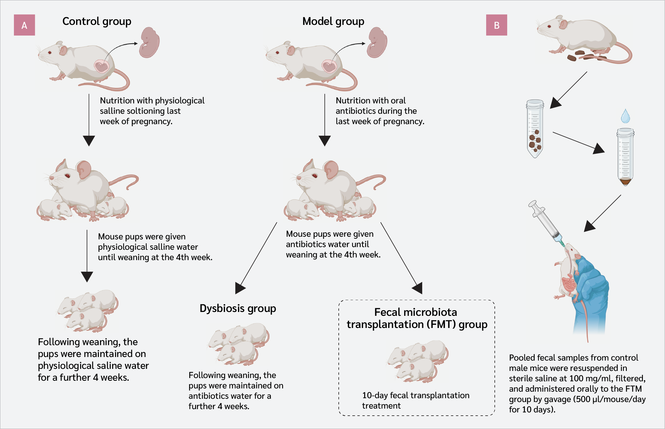
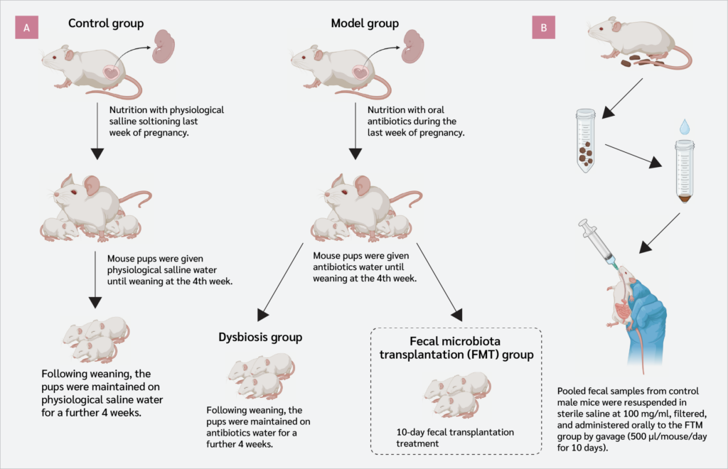
After inducing dysbiosis in the FMT group (i.e., after completion of the 1-week prenatal and 8-week postnatal antibiotic regimen), FMT was administered to the pups. Considering the antibiotic half-life, FMT was initiated 1 day after the last antibiotic dose. Healthy donor mice from the control group were used for FMT treatment (n=5). Fecal samples obtained from the control group mice were suspended and administered orally to the FMT group via gavage once daily for 10 days. Fecal samples were collected fresh daily. Fecal microbiota transplantation was not administered to the dysbiosis group (Figure 1B). All animals were housed in individually ventilated cage (IVC) (Tecniplast S.p.A., Buguggiate, Italy) systems with sterile feed and bedding. The experimental procedure used to induce microbial changes is summarized in Figure 1.
After microbial changes were completed, fecal samples were collected from all offspring groups to assess whether dysbiosis had developed and to evaluate the effect of FMT. Fecal pellets were collected into sterile, DNase- and RNase-free tubes free of DNA and RNA contamination. Samples from each group were pooled to create a single sample per group. Shotgun metagenomic sequencing was performed by Sapiens Genetic Tanı Inc. (İstanbul, Türkiye). In summary, library preparation was performed using the Illumina DNA Prep Kit (Illumina, San Diego, CA, USA,) according to the manufacturer's instructions, including DNA shearing, indexing, amplification, and purification. Library fragment distribution was assessed using the Agilent 2100 Bioanalyzer (Agilent Technologies, Santa Clara, CA, USA). Sequencing was performed on an Illumina NovaSeq 6000 platform (Illumina, San Diego, CA, USA) to obtain paired-end 150-bp reads.
Induction of Collagen-Induced Arthritis (CIA)
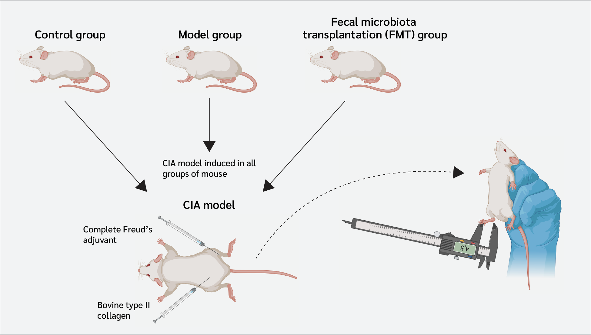
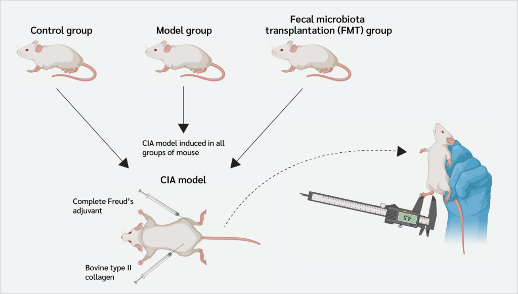
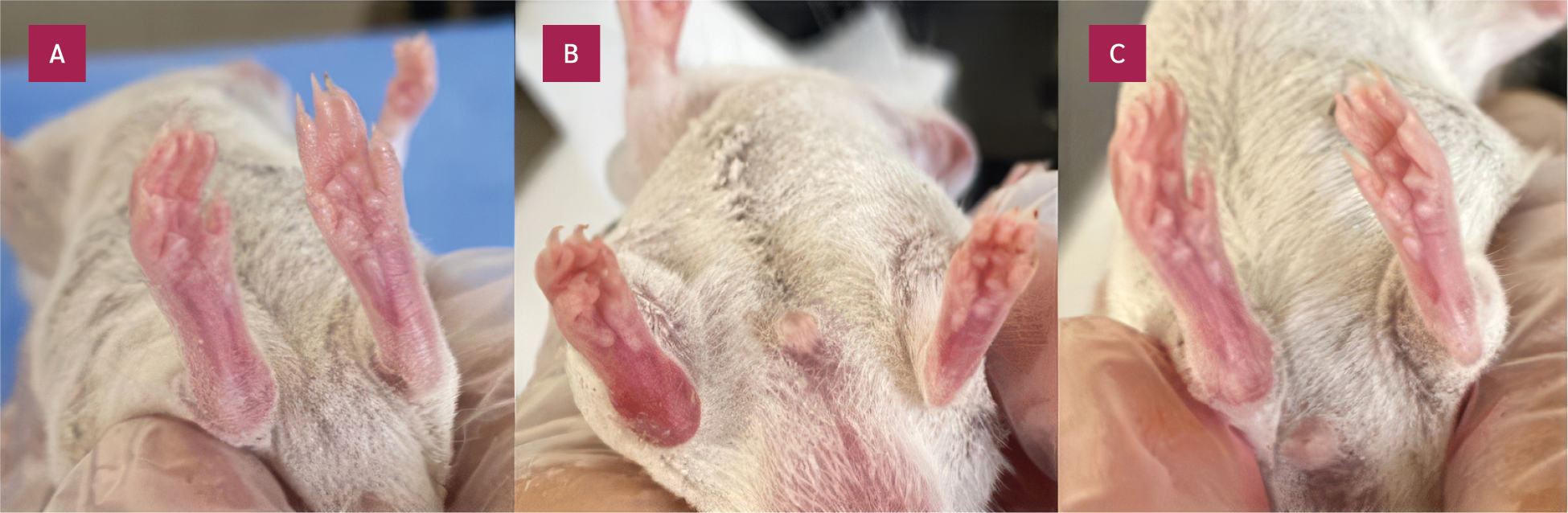
Following the alterations in microbial composition, CIA was initiated in male offspring across all groups, in accordance with a previously established method for BALB/c mice (19). To minimize potential variability associated with fluctuations in sex hormones and the estrus cycle in mice, only male offspring were included in the study. In summary, 10 µL of collagen emulsion (containing 100 µg bovine type II collagen; Sigma-Aldrich, St. Louis, MO, USA) was injected intra-articularly into the right hind leg (between the femur and tibia). At the same time, 100 µL of complete Freund's adjuvant (CFA; containing heat-killed Mycobacterium tuberculosis; Sigma-Aldrich, St. Louis, MO, USA) was injected subcutaneously. This immunization protocol was repeated 14 days later. Arthritis development was monitored for 21 days. The left hind leg of each animal that did not receive an injection served as a control (Figure 2).
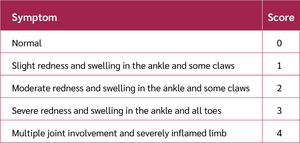
Clinical arthritis was assessed by measuring joint thickness with a digital caliper (Mitutoyo, Kawasaki, Japan) (Figure 2) and by scoring the severity of joint inflammation according to the clinical scoring system (Table 1). Body weight was measured every 3–4 days for 10 weeks.
Histopathological Analysis
At the end of the experiment (day 21), the mice were euthanized by cervical dislocation. Joint tissue samples were collected from the arthritic (right) and control (left) legs. The legs were disarticulated at the coxofemoral joint, skinned, and fixed in 10% neutral-buffered formalin (Sigma-Aldrich, St. Louis, MO, USA). Following fixation, bone decalcification was performed using 14% EDTA (pH 7.2; Sigma-Aldrich, St. Louis, MO, USA), and the solution was changed every three days. After decalcification, the joints were cut horizontally at the genu articulation region and embedded in paraffin following a standard tissue processing procedure (increasing alcohol concentrations, xylene, paraffin).
Sections 4 µm thick were cut from the paraffin blocks and stained with hematoxylin and eosin (Merck Millipore, Burlington, MA, USA) for examination under a light microscope (Olympus CX41 Phase Contrast & Darkfield Microscope, Tokyo, Japan). Histological scoring was performed using a standardized semi-quantitative scale (0-4), evaluating synovial hyperplasia, pannus formation, inflammatory cell infiltration of the synovial membrane, neovascularization, cartilage hypocellularity, and degenerative/necrotic changes accompanied by fissure formation. All sections were evaluated independently by two blinded investigators to ensure consistency and minimize observer bias.
Statistical Analysis
Statistical analyses were performed with GraphPad Prism 8 (GraphPad Software, San Diego, CA, USA). Continuous variables, including joint thickness values and body weight, were analyzed using one-way ANOVA followed by Tukey’s post hoc test. Ordinal data, including clinical arthritis scores and histopathological scores, were analyzed using the Kruskal–Wallis test followed by Dunn’s multiple comparison test. All data were presented as mean ± standard error of the mean. Statistical comparisons were performed using the independent samples t-test. A p-value <0.05 was considered statistically significant.
Results
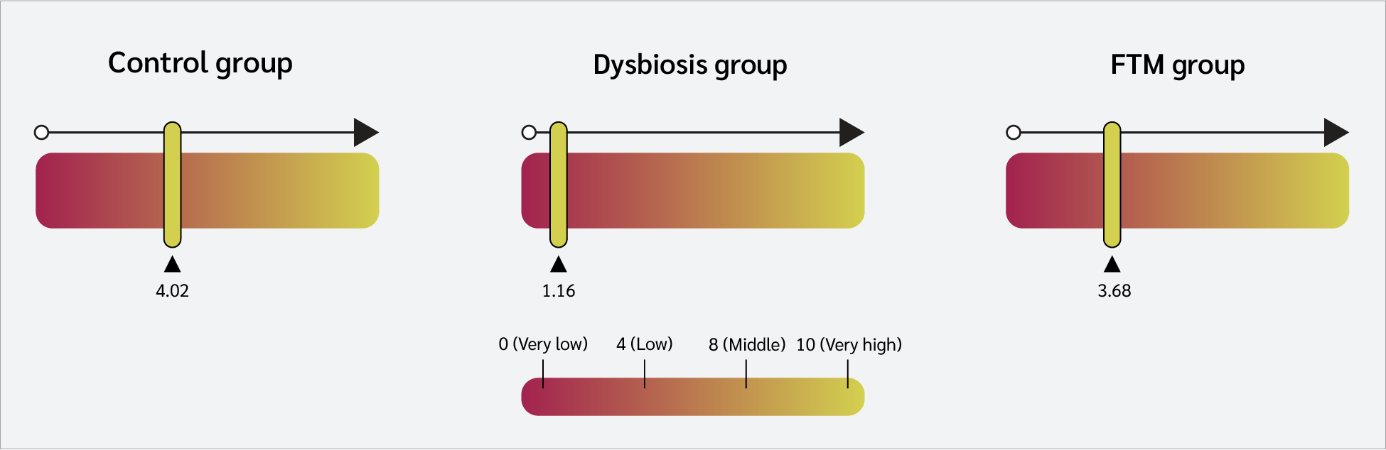
Shotgun metagenomic analysis of fecal samples collected prior to arthritis induction revealed significant changes in the gut microbiota. The Shannon index, an indicator of alpha diversity, was 4.02 in the control group and 1.16 in the dysbiosis group. These findings indicate that alpha diversity in the dysbiosis group was significantly reduced compared with that of the control group. The Shannon index of 3.68 in the FMT group indicated that FMT substantially restored this loss of diversity (Figure 3).
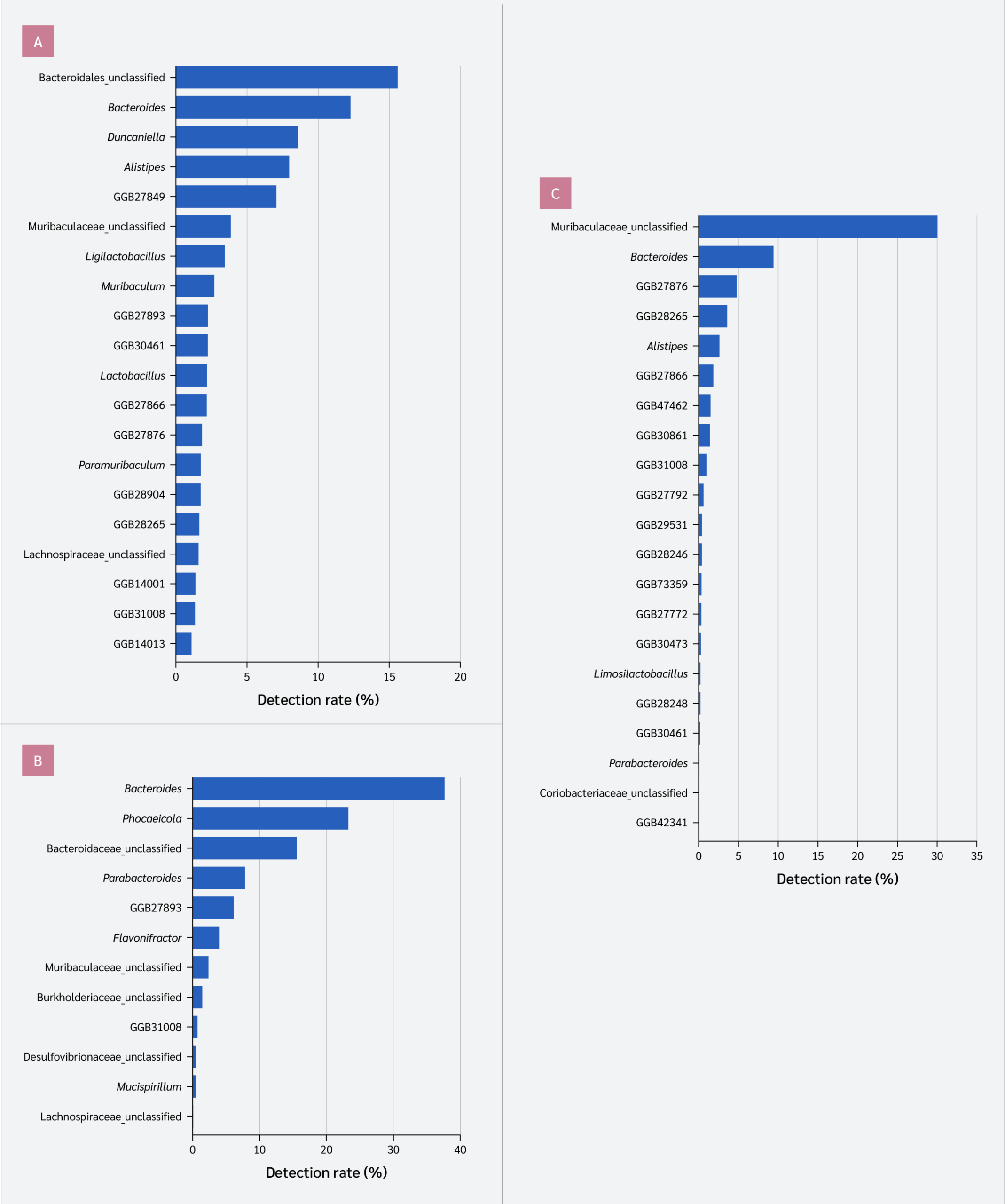
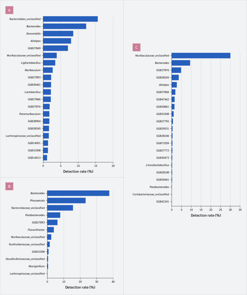
Genus-level bacterial distribution analysis showed that the microbial population was significantly reduced in the dysbiosis group. While 124 different bacterial genera were identified in the control group, this number dropped to 12 genera in the dysbiosis group. In the FMT group, bacterial diversity increased, with 109 genera identified, indicating restoration of the microbial diversity toward that observed in the control group (Figure 4).
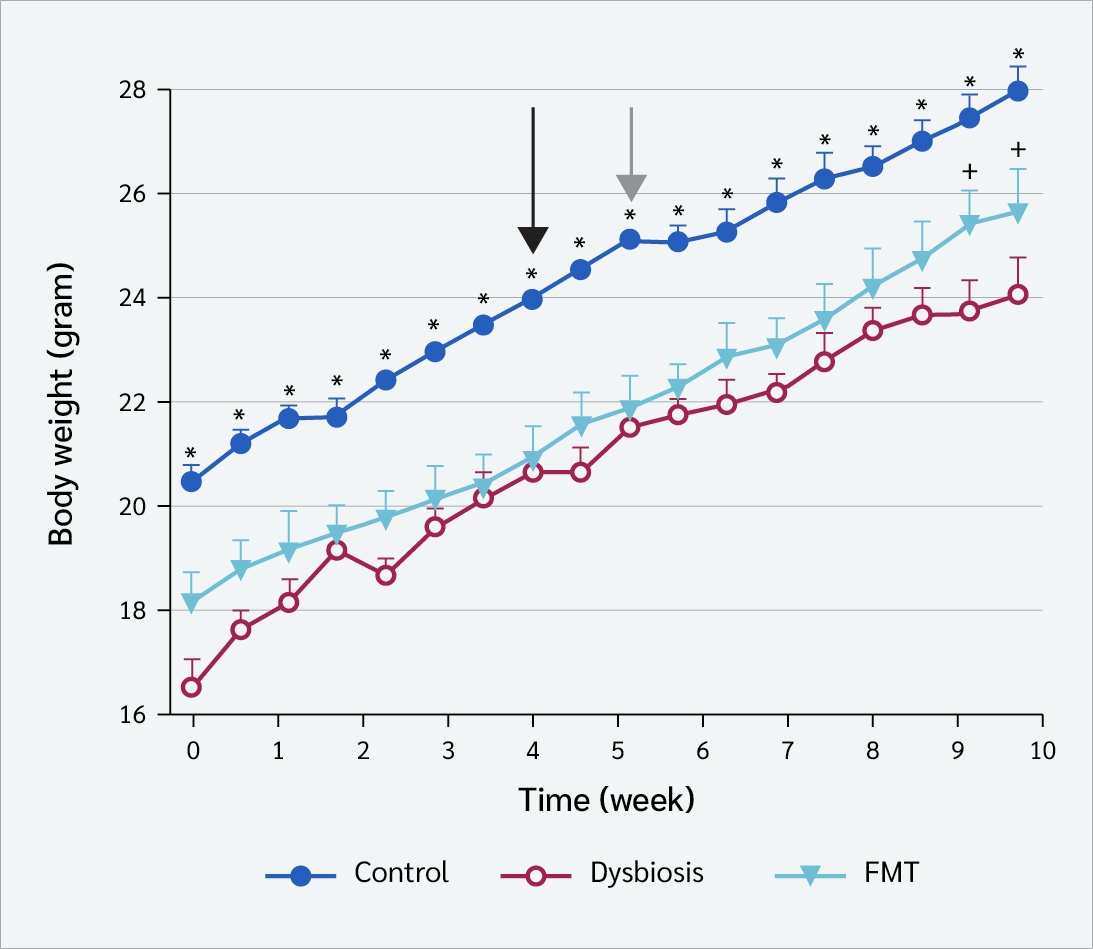
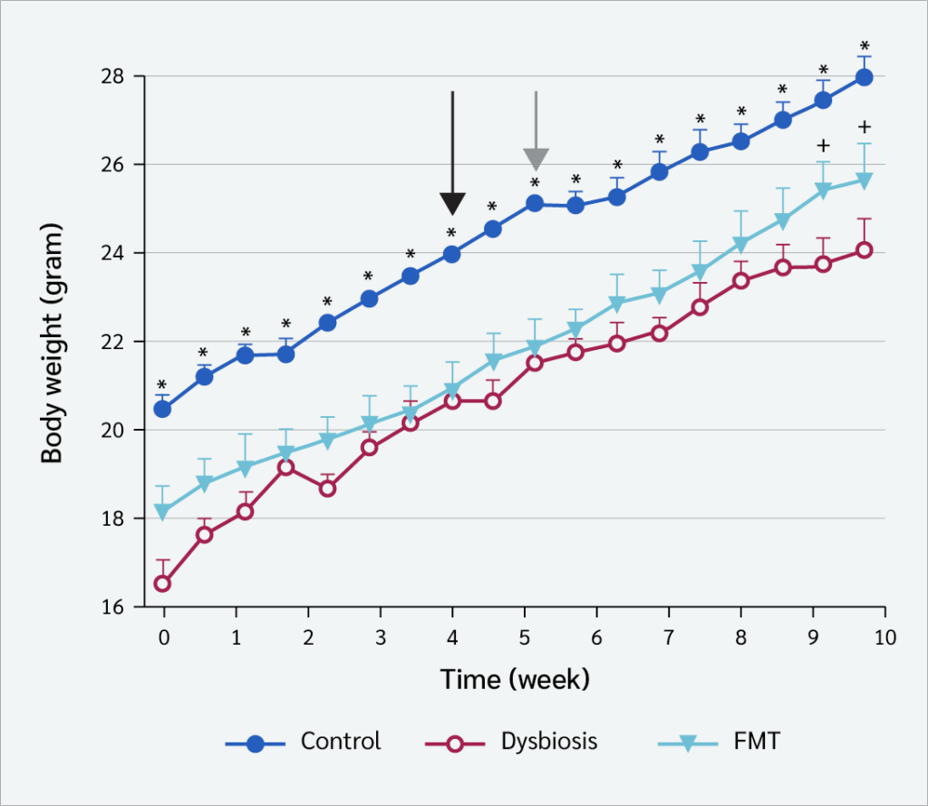
Maternal-initiated dysbiosis had a significant effect on growth. Animals in the dysbiosis and FMT groups had significantly lower body weights than those in the control group from the time of weaning (week 0). This difference persisted throughout the 4-week postnatal antibiotic administration period. After FMT treatment began, the FMT group showed an improving trend in body weight gain compared with the dysbiosis group. Following arthritis induction, body weight gain slowed in all groups as expected (Figure 5).

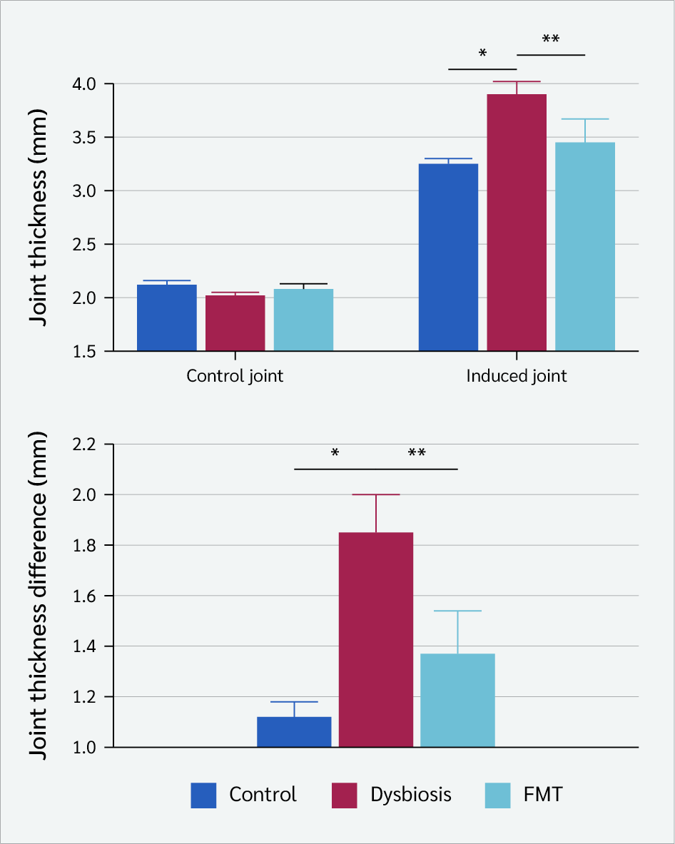
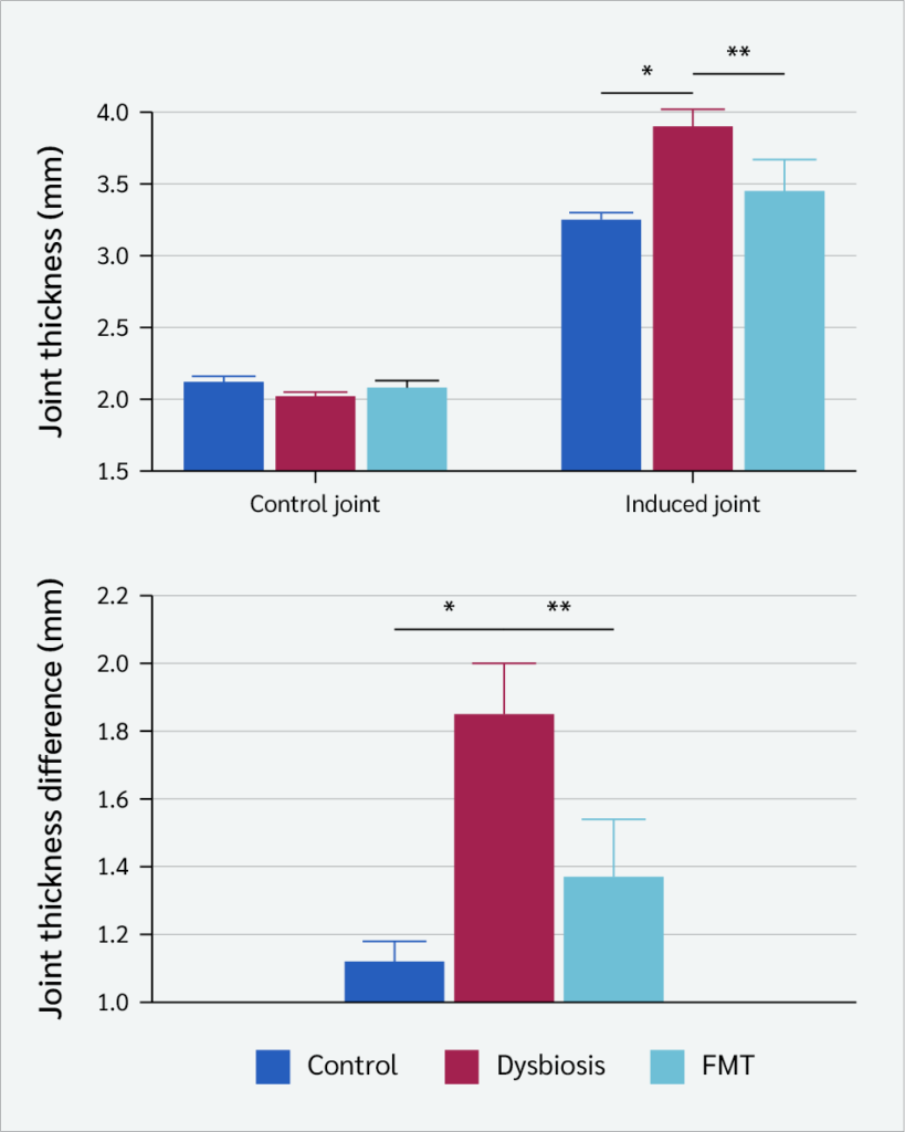
In the CIA model, joint thickness was significantly higher in the dysbiosis group than in the control group (p=0.0028). Joint thickness was significantly lower in the FMT group than in the dysbiosis group (p=0.046) (Figure 6).
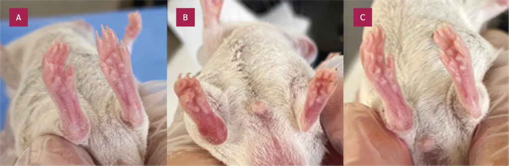
When the difference in thickness between the arthritic and control joints was calculated, it became evident that dysbiosis increased the severity of arthritis, while FMT partially prevented this increase (Figure 6). Clinical arthritis scores showed no statistically significant differences between the groups (Table 2). Images of the joints in the experimental CIA model are shown in Figure 7.
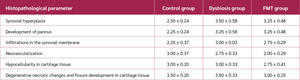
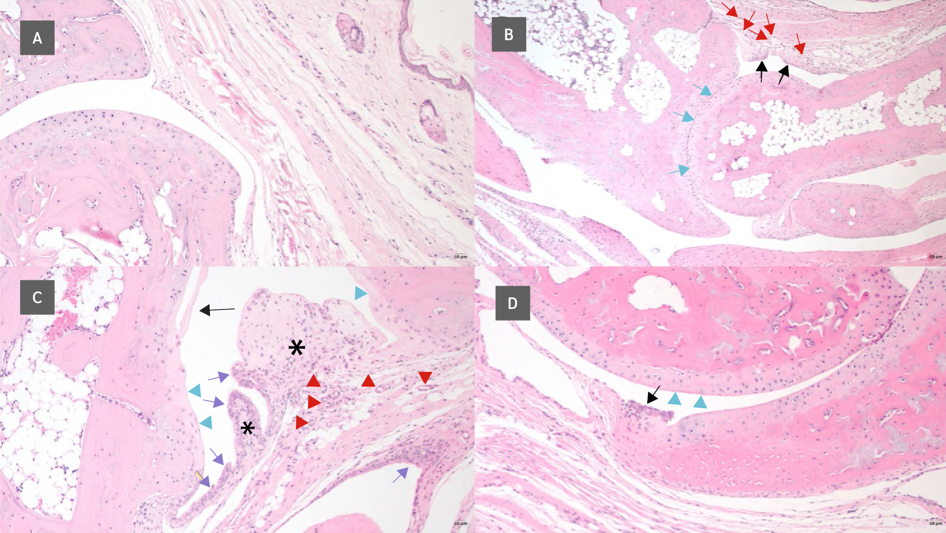
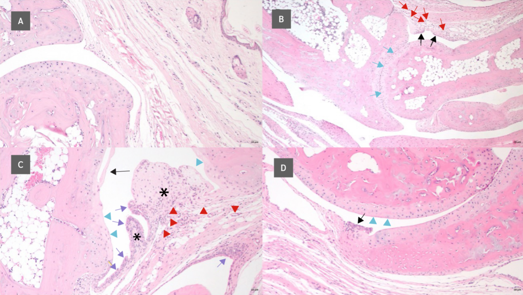
Histopathological examination of joint tissues revealed that dysbiosis exacerbated arthritis-related tissue damage. In the dysbiosis group, more severe synovial hyperplasia, pannus formation, cartilage erosion, and near-complete loss of the joint space were observed compared with the control group. In the FMT group, the severity of these pathological changes was reduced compared with the dysbiosis group (Figure 8). Histopathological scoring confirmed higher damage scores for parameters such as synovial hyperplasia and pannus formation in the dysbiosis group, while demonstrating that FMT reduced this damage (Table 3).
Discussion
This study examined the impact of antibiotic-induced dysbiosis during the maternal period on the severity of CIA in offspring and the possible therapeutic effects of FMT.
In this study, we demonstrated that maternal dysbiosis reduced gut microbial diversity, led to reduced body weight gain in offspring, and increased arthritis pathology. Importantly, FMT partially restored microbial diversity and attenuated arthritis severity. The most important finding of our study is that maternal dysbiosis exacerbates the pathogenesis of arthritis.
Our findings suggest that maternal dysbiosis was associated with reduced body weight gain in male offspring, which may indicate a potential impact on early developmental parameters. However, further studies are needed to draw more definitive conclusions regarding developmental outcomes. This result is consistent with previous research showing an association between antibiotic use and an increased prevalence of RA (20).
Similarly, an increase in arthritis severity has also been reported in experimental models where antibiotics were administered postpartum (21,22). However, our study makes a significant contribution to the literature by showing that the risk occurs not only in the postnatal period but also in the maternal period. This finding highlights the critical role of the gut microbiota in the early development of the immune system.
Our microbiota analyses showed that the combination of amoxicillin and vancomycin caused significant dysbiosis and that FMT was able to substantially restore microbial diversity. We also found that the combination of amoxicillin and vancomycin led to a significant decrease in the Firmicutes/Bacteroidetes ratio within the bacterial population. This finding can be explained by the well-documented activity of vancomycin against Gram-positive bacteria (23).
Our analysis further demonstrated that the number of bacterial genera increased after FMT. The increase in the number of bacterial genera supports the ability of FMT to restore microbial diversity, and this finding is consistent with previous studies (24,25). For example, a clinical study conducted in 2018 (24) demonstrated that FMT increases gut microbial diversity in humans and is a reliable method. Similarly, another study (25) reported an increase in the microbial population and Shannon index following FMT, consistent with our findings. Numerous animal models have also demonstrated that FMT can reshape the gut microbiome, supporting our results (26,27). According to our results, it can be considered that dysbiosis may disrupt the integrity of the intestinal epithelial barrier, increasing the passage of microbial products (such as lipopolysaccharide [LPS]) into the systemic circulation, thereby triggering the production of pro-inflammatory cytokines (tumor necrosis factor-alpha [TNF-α], interleukin-6 [IL-6], and interleukin-17 [IL-17]) and exacerbating joint inflammation.
Additionally, the decrease in Lachnospiraceae observed in the dysbiosis group is significant because this family is a major producer of short-chain fatty acids (e.g., butyrate) with anti-inflammatory effects. The decrease in these metabolites may have contributed to impaired immune tolerance and enhanced autoimmune responses. The protective effect of FMT is likely related to its ability to restore these beneficial bacteria, thereby promoting the production of immunomodulatory metabolites.
There are some limitations to our study. First, the number of male offspring obtained after birth was relatively low. Second, to explain the mechanism more clearly, measurements of intestinal permeability, analysis of serum cytokine levels, and examination of immune cell subpopulations in the lamina propria or spleen (such as the regulatory T cell [Treg]/T helper 17 [Th17] balance) would be required. Additionally, metabolomic analyses would further improve our understanding of the functional capacity of the microbiota alterations.
Another limitation of our study relates to the metagenomic sequencing methodology. Due to budget constraints, fecal samples from five animals in each experimental group were pooled together, creating a single composite sample for sequencing analysis for each group. While this approach provided valuable insights into the microbial community structure at the group level and successfully demonstrated the profound effects of antibiotic-induced dysbiosis and FMT-mediated restoration, it prevented us from assessing inter-individual differences in gut microbiota composition. Consequently, we were unable to evaluate the heterogeneity of microbial responses within treatment groups or perform statistical analyses on alpha- and beta-diversity metrics at the individual level. Future studies with larger sample sizes and individual sequencing will be valuable for characterizing the full spectrum of microbial diversity and elucidating individual differences in response to dysbiosis and FMT treatment.
In conclusion, this study demonstrated that maternal-derived gut dysbiosis can influence the severity of CIA development in mice. It has been shown that FMT treatment holds therapeutic potential to reduce this severity. Our findings highlight the importance of the microbiome in the development and severity of autoimmune diseases and suggest that microbiota-focused treatment approaches may represent a promising preventive or therapeutic approach.
Ethical Approval
This experimental study was approved by the Bursa Uludağ University Animal Experiments Local Ethics Committee on April 11, 2023 with decision number 2023-06/05.
Informed Consent
Not applicable.
Peer-review
Externally peer-reviewed
Author Contributions
Concept – H.B.O., M.Y., D.Y.E.; Design – H.B.O., M.Y., D.Y.E.; Supervision – H.B.O., M.Y., D.Y.E.; Funding – H.B.O.; Materials – H.B.O., D.Y.E., E.Y.; Data Collection and/or Processing – F.D., G.A., E.Y., M.A., A.A.; Analysis and/or Interpretation – F.D., G.A., E.Y., M.A., A.A., M.Y.; Literature Review – F.D., H.B.O., M.Y., D.Y.E., G.A.; Writing – F.D., H.B.O., M.Y., D.Y.E., A.A.; Critical Reviews – H.B.O., M.Y., D.Y.E.
Conflict of Interest
The authors declared that they have no conflict of interest.
Financial Disclosure:
This study was supported by the Bursa Uludağ University Research Projects Coordination Office under Grant Nos. TYL-2023-1567 and TAY-2022-601.
AI Statement
During the preparation of this manuscript, the authors used Writefull to improve the language, grammar, and readability of the manuscript. The authors reviewed and edited the generated output as necessary and take full responsibility for the content of this publication.
Scientific Presentation
This study was presented as an oral presentation at the Immuno-Rheumatology Symposium, held in Antalya, Türkiye, on February 20–25, 2025.
Acknowledgment
The authors thank the Bursa Uludağ University Research Projects Coordination Office (BUÜ BAP Unit) for its financial support.
References
Gensollen T, Iyer SS, Kasper DL, Blumberg RS. How colonization by microbiota in early life shapes the immune system. Science. 2016;352(6285):539–44. [CrossRef]
Wu HJ, Wu E. The role of gut microbiota in immune homeostasis and autoimmunity. Gut Microbes. 2012;3(1):4–14. [CrossRef]
Sender R, Fuchs S, Milo R. Revised estimates for the number of human and bacteria cells in the body. PLoS Biol. 2016;14(8):e1002533. [CrossRef]
Groschwitz KR, Hogan SP. Intestinal barrier function: molecular regulation and disease pathogenesis. J Allergy Clin Immunol. 2009;124(1):3–20; quiz 21–2. [CrossRef]
Jernberg C, Löfmark S, Edlund C, Jansson JK. Long-term impacts of antibiotic exposure on the human intestinal microbiota. Microbiology (Reading). 2010;156(Pt 11):3216–23. [CrossRef]
Wampach L, Heintz-Buschart A, Fritz JV, Ramiro-Garcia J, Habier J, Herold M, et al. Birth mode is associated with earliest strain-conferred gut microbiome functions and immunostimulatory potential. Nat Commun. 2018;9(1):5091. [CrossRef]
Francino MP. Antibiotics and the human gut microbiome: dysbioses and accumulation of resistances. Front Microbiol. 2016;6:1543. [CrossRef]
Scher JU, Sczesnak A, Longman RS, Segata N, Ubeda C, Bielski C, et al. Expansion of intestinal Prevotella copri correlates with enhanced susceptibility to arthritis. Elife. 2013;2:e01202. [CrossRef]
Maeda Y, Takeda K. Role of gut microbiota in rheumatoid arthritis. J Clin Med. 2017;6(6):60. [CrossRef]
Horta-Baas G, Romero-Figueroa MDS, Montiel-Jarquín AJ, Pizano-Zárate ML, García-Mena J, Ramírez-Durán N. Intestinal dysbiosis and rheumatoid arthritis: a link between gut microbiota and the pathogenesis of rheumatoid arthritis. J Immunol Res. 2017;2017:4835189. [CrossRef]
Romero-Figueroa MDS, Ramírez-Durán N, Montiel-Jarquín AJ, Horta-Baas G. Gut-joint axis: gut dysbiosis can contribute to the onset of rheumatoid arthritis via multiple pathways. Front Cell Infect Microbiol. 2023;13:1092118. [CrossRef]
Ivanov II, Atarashi K, Manel N, Brodie EL, Shima T, Karaoz U, et al. Induction of intestinal Th17 cells by segmented filamentous bacteria. Cell. 2009;139(3):485–98. [CrossRef]
Scher JU, Abramson SB. Periodontal disease, Porphyromonas gingivalis, and rheumatoid arthritis: what triggers autoimmunity and clinical disease? Arthritis Res Ther. 2013;15(5):122. [CrossRef]
Schepici G, Silvestro S, Bramanti P, Mazzon E. The gut microbiota in multiple sclerosis: an overview of clinical trials. Cell Transplant. 2019 Dec;28(12):1507–27. [CrossRef]
Kishikawa T, Maeda Y, Nii T, Okamoto A, Katoh Y, Ohta K et al. Metagenome-wide association study of gut microbiome in rheumatoid arthritis identifies novel microbial biomarkers and reveals immune cell cross-talk. Annals of the Rheumatic Diseases. 2023;82(10):1279–87. [CrossRef]
van Nood E, Vrieze A, Nieuwdorp M, Fuentes S, Zoetendal EG, de Vos WM, et al. Duodenal infusion of donor feces for recurrent Clostridium difficile. N Engl J Med. 2013;368(5):407–15. [CrossRef]
Ray R, Hack SA, Vij AK, Gbenla KI, Khatri S, Aravind Rongali D, et al. Efficacy of fecal microbiota transplantation (FMT) versus standard antibiotic therapy in recurrent Clostridioides difficile (CDI/rCDI) infection: a systematic review and meta-analysis. Cureus. 2025;17(8):e90614. [CrossRef]
Belvoncikova P, Maronek M, Gardlik R. Gut dysbiosis and fecal microbiota transplantation in autoimmune diseases. Int J Mol Sci. 2022;23(18):10729. [CrossRef]
Güvenç G, Karaçay M, Akkoç A, Çiftçi K, Oral HB, Yalçın M. A New protocol for collagen-induced local arthritis model in Balb/c Mice. Turk J Immunol. 2018;6(1):30–6. [CrossRef]
Sultan AA, Mallen C, Muller S, Hider S, Scott I, Helliwell T, Hall LJ. Antibiotic use and the risk of rheumatoid arthritis: a population-based case-control study. BMC Med. 2019;17(1):154. [CrossRef]
Dorożyńska I, Majewska-Szczepanik M, Marcińska K, Szczepanik M. Partial depletion of natural gut flora by antibiotic aggravates collagen induced arthritis (CIA) in mice. Pharmacol Rep. 2014 Apr;66(2):250–5. Erratum in: Pharmacol Rep. 2017;69(3):586. [CrossRef]
Yang S, Chen H, Wei B, Xiang M, Hu Z, Peng Z, et al. Clindamycin administration increases the incidence of collagen-induced arthritis in mice through the prolonged impact of gut immunity. Inflammation. 2018;41(5):1900–11. [CrossRef]
Tulstrup MV, Christensen EG, Carvalho V, Linninge C, Ahrné S, Højberg O, et al. Antibiotic treatment affects intestinal permeability and gut microbial composition in wistar rats dependent on antibiotic class. PLoS One. 2015;10(12):e0144854. [CrossRef]
DeFilipp Z, Peled JU, Li S, Mahabamunuge J, Dagher Z, Slingerland AE, et al. Third-party fecal microbiota transplantation following allo-HCT reconstitutes microbiome diversity. Blood Adv. 2018;2(7):745–53. [CrossRef]
Seekatz AM, Aas J, Gessert CE, Rubin TA, Saman DM, Bakken JS, et al. Recovery of the gut microbiome following fecal microbiota transplantation. mBio. 2014;5(3):e00893–14. [CrossRef]
Wang J, Zhou X, Li X, Guo W, Zhu Q, Zhu B, et al. Fecal microbiota transplantation alters the outcome of hepatitis B virus infection in mice. Front Cell Infect Microbiol. 2022;12:844132. [CrossRef]
Tian Z, Liu J, Liao M, Li W, Zou J, Han X, et al. Beneficial effects of fecal microbiota transplantation on ulcerative colitis in mice. Dig Dis Sci. 2016;61(8):2262–71. [CrossRef]
VOLUME
,
ISSUE
Correspondence
Received
Accepted
Published
Suggested Citation
DOI
License

