Comparative Evaluation of Magnetic Cell Separation Systems Based on Cell Recovery and CD14 mRNA Enrichment


Abstract
Objective:
Efficient enrichment of CD14-expressing cell fractions is important for downstream applications such as in vitro differentiation and molecular analyses. However, selecting an optimal magnetic separation method can be challenging, particularly when both cell yield and enrichment efficiency must be considered. To address this challenge, we compared two magnetic separation platforms, MACS™ and MojoSort™, that use anti-CD14 antibody-conjugated beads for positive selection, focusing on cell recovery and CD14 mRNA enrichment.
Materials and Methods:
Peripheral blood mononuclear cells (PBMCs) were isolated from eight healthy pediatric donors using Ficoll gradient centrifugation. The performance of two immunomagnetic cell isolation methods utilizing antibody-conjugated magnetic particles, differing in separation format (column-based vs. column-free) was evaluated based on viable cell recovery and relative CD14 gene expression levels measured by RT-qPCR across input and output fractions, including PBMCs, CD14-enriched fractions, and flow-through fractions.
Results:
Column-based method demonstrated higher cell recovery and yielded fractions with significantly greater CD14 mRNA expression compared to column-free system. Both systems produced fractions with increased CD14transcript levels relative to input PBMCs and corresponding flow-through fractions. Cells derived from both methods exhibited comparable expression of macrophage-associated genes following in vitro differentiation.
Conclusion:
Under the tested conditions, Column-based separation system provided higher cell recovery and stronger enrichment of CD14 mRNA expression than the column-free system. These findings highlight differences in enrichment efficiency at the transcript level, while both systems generated cell fractions suitable for downstream molecular and functional applications.
Keywords:
Magnetic, cell, separation CD14, mRNA, enrichment column-free, magnetic, separation, system column-based, magnetic, separation, system RT-qPCRIntroduction
Magnetic cell sorting is a rapid and reliable technique commonly used to isolate specific cell types from blood or tissue samples. Anti-CD14 magnetic beads are commonly employed in positive selection strategies, as CD14 is predominantly expressed in monocyte-lineage cells, including monocytes, macrophages, and certain dendritic cell subsets (1–5).
The choice of cell separation strategy can influence not only cell recovery but also downstream cellular behavior, including differentiation potential. Accordingly, obtaining viable and sufficiently enriched cell fractions is critical for applications such as in vitro differentiation and molecular analyses. However, achieving efficient enrichment can be particularly challenging in pediatric settings, where limited blood volume and low peripheral blood mononuclear cell (PBMC) counts often restrict the number of cells available for downstream applications. These limitations may result in insufficient recovery of CD14-expressing cell fractions for use in cell culture or nucleic acid–based analyses.
In addition, monocyte-lineage cells are relatively sensitive to handling, and the multi-step procedures required for PBMC isolation and subsequent magnetic separation can introduce variability in both yield and enrichment efficiency.
Several magnetic-bead-based cell separation systems are currently available, including column-based system, (MACS™; Miltenyi Biotec, Bergisch Gladbach, Germany) and column-free system (MojoSort™; BioLegend, San Diego, CA, USA). Among these, magnetic-activated cell column-based sorting system (MACS™) is one of the most widely used systems and relies on direct magnetic labeling with antibody-conjugated microbeads. In this system, labeled cells are retained within a magnetic column, while unlabeled cells are removed, followed by elution of the retained fraction (2,6).
In contrast, the column-free system (MojoSort™) system uses indirect magnetic labeling of cells prior to separation in a magnetic field, first with the primary antibody, then with magnetic streptavidin nanobeads (7–9). Although both approaches have been reported to be effective, differences in labeling strategy and workflow may influence cell recovery and enrichment efficiency.
In the present study, we compared two magnetic bead–based separation systems, focusing on post-separation cell viability and the enrichment of CD14 mRNA expression. Separation performance was assessed by measuring viable cell yield through cell counting and relative CD14 transcript levels across input and output fractions using reverse transcription quantitative polymerase chain reaction (RT-qPCR).
In addition, the functional potential of the enriched fractions was assessed through in vitro macrophage differentiation assays. Overall, this study aimed to provide a practical and reproducible framework for optimizing magnetic separation workflows, particularly in settings where sample availability is limited, and evaluation is primarily based on transcript-level analyses.
Materials and Methods
Sample Collection
Peripheral blood samples were obtained from healthy pediatric donors (n=8) in accordance with an approved institutional ethics committee protocol and following informed consent from parents or legal guardians. All donors were clinically evaluated prior to inclusion, had no history of rheumatic or inflammatory diseases, and were not receiving any medication at the time of sampling.
In addition, inflammatory markers, including C-reactive protein (CRP) and erythrocyte sedimentation rate (ESR), were within normal reference ranges, confirming the absence of active systemic inflammation. Blood samples were collected into ethylenediaminetetraacetic acid (EDTA)-coated tubes (16 × 100 mm) under sterile conditions and processed on the same day to preserve cell integrity.
Peripheral blood samples were collected from each donor into two 8 mL EDTA tubes. For the EDTA comparison experiments, blood from one tube was divided into three equal portions, with approximately 2.5 mL allocated to each experimental condition. For the magnetic separation experiments, PBMCs isolated from the other 8 mL blood sample were divided into two equal fractions and subjected to MojoSort™ and MACS™ separation protocols, respectively.
All experimental procedures were conducted in accordance with relevant guidelines and regulations. Demographic and clinical characteristics of the donors, including age, sex, body mass index (BMI), CRP, and ESR values, are provided in Table S1.
Isolation of Peripheral Blood Mononuclear Cells
Equal volumes of peripheral blood were mixed with Dulbecco's phosphate-buffered saline without calcium and magnesium, or with DPBS supplemented with 1–2 mM EDTA (0.5 M stock solution, pH 8.0, RNase-free; Thermo Scientific™, Waltham, MA, USA) in 15- or 50- mL Falcon tubes. The blood-DPBS mixture was layered onto the Ficoll-Paque™ Plus (density 1.077 g/mL; GE Healthcare, Chicago, IL, USA) by placing the pipette tip against the inner wall of the tube.
The samples were centrifuged at 400 × g for 40 minutes, with the brake off, at 18-20°C. The buffy coat layer, containing PBMCs, was drawn between the plasma and Ficoll-Paque layers and washed twice with three volumes of DPBS (with or without EDTA) by centrifugation at 450 × g for 10 minutes.
Cell counting was performed using a hemocytometer using 0.4% Gibco™ Trypan Blue Solution (Thermo Fisher Scientific, Waltham, MA, USA). All procedures involving cells were conducted in a biosafety level 2 (BSL-2) cell culture hood.
Technical challenges encountered during the experimental workflow, along with corresponding troubleshooting strategies, are summarized in Table S2. Peripheral blood mononuclear cells isolated from each donor were divided into equal fractions (5 × 10⁶ input cells per condition) and subjected to two magnetic separation systems in parallel, allowing paired comparison within the same donor. Additionally, PBMCs isolated using DPBS without EDTA were subsequently used for magnetic sorting experiments.
Magnetic Cell Separation Using Miltenyi Biotec MACS™
Peripheral blood mononuclear cells from each sample were first passed through a 40-µm cell strainer (Corning, Corning, NY, USA) to obtain a single-cell suspension prior to magnetic labeling. Following cell counting,
1 × 10⁷ PBMCs were divided into two aliquots, with
5 × 10⁶ input cells allocated to each separation method, and centrifuged at 300 × g for 5 – 10 minutes at 4°C.
The supernatant was completely aspirated, and 80 μL ice-cold MACS buffer (prepared with 0.5% bovine serum albumin [BSA], 2 mM EDTA, and DPBS without Ca++ and Mg++, and filtered using the Stericup® Quick Release Vacuum Filtration System; MilliporeSigma, Burlington, MA, USA) was added to the cell pellet. The suspension was then transferred into a 5-mL polypropylene round-bottom tube (Corning, Corning, NY, USA).
To minimize non-specific antibody binding, Human TruStain FcX™ Fc Receptor Blocking Solution (BioLegend, San Diego, CA, USA; cat. no. 422301) was added to the cell suspension. After 10 min of incubation at room temperature, 20 μL MACS™ CD14 MicroBeads (Miltenyi Biotec, Bergisch Gladbach, Germany; cat. no. 130-050-201) was added, and the mixture was incubated for 15 minutes on ice with shaking.
The volume was then adjusted to 3 mL with MACS buffer, and the cells were gently mixed and centrifuged at 300 × g for 5 minutes at 4°C. LS columns (Miltenyi Biotec; cat. no. 130-042-401) were rinsed with 3 mL MACS buffer, and the cell pellet was resuspended in 500 µL of MACS buffer.
The cell suspension was applied onto the column placed in a MidiMACS™ Separator (Miltenyi Biotec; cat. no. 130-042-302), and the flow-through containing unlabeled cells was collected. After all the cell mixture left the reservoir, the column was washed twice with 3 mL MACS buffer.
The column was then removed from the magnetic field, and the retained CD14-positive cells were immediately flushed out with 5 mL MACS buffer by forcefully pushing the plunger. Both fractions were spun at 300 × g for 10 minutes at 4°C.
Cell counting was performed using trypan blue solution, and the cells were separated for either nucleic acid isolation or in vitro macrophage differentiation.
Magnetic Cell Separation Using BioLegend MojoSort™
In parallel with the MACS™ protocol, 5 × 10⁶ PBMCs were transferred into a 15-mL Falcon tube and centrifuged at 300 × g for 5–10 minutes at 4°C. The supernatant was completely aspirated, and 100 μL ice-cold MACS buffer was added to the cell pellet. The suspension was then transferred into a 5-mL polypropylene round-bottom tube.
To reduce non-specific antibody binding, 5 μL of Human TruStain FcX™ Fc Receptor Blocking Solution (BioLegend, San Diego, CA, USA; cat. no. 422301) was added, and the cells were incubated at room temperature for 10 minutes.
Subsequently, 10 μL of MojoSort™ Biotin Anti-Human CD14 Antibody (BioLegend; cat. no. 480048) was added, and the cells were incubated for 15 minutes. This was followed by the addition of 10 μL of MojoSort™ Streptavidin Nanobeads (BioLegend), with an additional 15 minutes of incubation on ice with shaking.
The total volume was then adjusted to 4 mL with MACS buffer, and the cells were centrifuged at 300 × g for 5 minutes at 4°C. The pellet was resuspended in 2.5 mL of MACS buffer, and the tube was placed in the MojoSort™ Magnet (BioLegend; cat. no. 480019) for 5 minutes at room temperature.
Without removing the tube from the magnetic separator, the supernatant containing the unlabeled cells (remaining in the middle of the tube) was carefully decanted into a clean tube. The magnetic separation step was repeated twice to improve purity.
After the final separation, the tube was removed from the magnet, and the collected putative monocyte-enriched fraction was resuspended in 3 mL MACS buffer. Both fractions were centrifuged at 300 × g for 10 minutes at 4°C, then counted using trypan blue solution.
The resulting cells were subsequently used for nucleic acid isolation or in vitro macrophage differentiation assays. Non-target fractions obtained from both methods were used as labeling controls.
Specifically, the number of PBMCs processed was standardized to 5 × 10⁶ cells for each method, and bead volumes were applied according to the manufacturer’s recommended ratios for this input cell number.
Assessment of Cell Viability and Separation Yield
For each donor, isolated PBMCs were split into two equal fractions and processed in parallel using the MACS™ and MojoSort™ separation kits. Peripheral blood mononuclear cells, magnetically selected fractions, and the corresponding flow-through cells were counted using trypan blue exclusion.
Cell viability and separation yield were calculated as follows:
Post-sort cell viability (%) = (Number of viable cells after sorting / Total number of recovered cells after sorting) × 100
Separation yield (%) = (Number of cells obtained in the magnetically selected CD14-enriched fraction after sorting / Input PBMCs) × 100
RNA Extraction and Quantitative PCR
Total RNA was extracted from equal numbers of cells derived from PBMCs, magnetically selected fractions, and corresponding flow-through cells using the PureLink™ RNA Mini Kit (Invitrogen™, Thermo Fisher Scientific, Waltham, MA, USA) according to the manufacturer’s instructions.
RNA concentration and purity were assessed spectrophotometrically using the NanoDrop™ One Microvolume Ultraviolet-Visible (UV-Vis) Spectrophotometer (Thermo Scientific™, Thermo Fisher Scientific, Waltham, MA, USA).
Complementary DNA (cDNA) was synthesized from equal input amounts of total RNA (200 ng) using the OneScript Plus cDNA Synthesis Kit (Applied Biological Materials Inc., Richmond, BC, Canada) following the manufacturer’s protocol.
RT-qPCR was performed using BlasTaq™ 2X qPCR MasterMix (Applied Biological Materials Inc., Richmond, BC, Canada) on a LightCycler® 480 Real-Time PCR System (Roche Diagnostics, Basel, Switzerland). Each reaction was performed in technical replicates using gene-specific primers (Table S3) and standardized amounts of cDNA.
Relative gene expression levels were calculated using the 2⁻ΔCt method, after normalization to an internal housekeeping gene glyceraldehyde-3-phosphate dehydrogenase (GAPDH) (10).
In Vitro Macrophage Differentiation
Cells from the magnetically selected fraction were seeded at a density of 3 × 10⁴ cells/cm² in 12-well tissue culture-treated plates containing RPMI-1640 medium (Gibco™, Thermo Fisher Scientific, Waltham, MA, USA; cat. no. 21875034) supplemented with 2 mM GlutaMAX™ (Gibco™, Thermo Fisher Scientific, Waltham, MA, USA), 10% heat-inactivated standardized commercial newborn calf serum (NBCS; Gibco™, Thermo Fisher Scientific, Waltham, MA, USA), 1% NEAA (Life Technologies), 1% sodium pyruvate (Gibco™, Thermo Fisher Scientific, Waltham, MA, USA), and 500 U/mL of penicillin-streptomycin (Gibco™, Thermo Fisher Scientific, Waltham, MA, USA).
To induce macrophage differentiation, the culture medium was further supplemented with 25 ng/mL recombinant human granulocyte-macrophage colony-stimulating factor (GM-CSF; R&D Systems, Minneapolis, MN, USA; cat. no. 215-GM-010) and 50 ng/mL macrophage colony-stimulating factor (M-CSF; R&D Systems, Minneapolis, MN, USA; cat. no. 216-MCC-010).
Cells were maintained under standard culture conditions (37°C, 5% CO₂) for 7 days, with partial medium replacement performed at regular intervals. At the end of the differentiation period, adherent macrophages were gently washed and either collected or directly used for downstream functional and molecular analyses.
Cell morphology was routinely examined using an inverted light microscope (Axio Vert.A1, ZEISS, Oberkochen, Germany).
Statistical Analysis
Statistical analysis was performed using IBM SPSS Statistics for Windows, version 29.0 (IBM Corp., Armonk, NY, USA). Data were shown as mean ± standard deviation (SD).
For comparisons between the two isolation methods, paired statistical tests were performed, as measurements obtained from the same eight donors constitute dependent (matched) quantitative data. All comparisons were conducted using paired samples derived from the same donor to eliminate inter-individual variability.
Normality of the paired differences was assessed using the Shapiro-Wilk test. When data were normally distributed, group comparisons were conducted using a paired t-test; otherwise, the Wilcoxon signed-rank test was applied.
A p-value < 0.05 was considered statistically significant.
Results
Optimizing the Working Conditions of PBMC Isolation
In this protocol comparison and optimization study, we first evaluated several critical parameters that can be decisive for efficient PBMC isolation. One key parameter examined was working temperature. Although standard protocols recommend maintaining all consumables used for PBMC isolation at room temperature (18–22°C), lower temperatures are often preferred to preserve cell viability during long isolation periods.
When cold DPBS was used, and centrifugation was performed at 4°C, the resulting buffy coat appeared clumpy (Figure S1a) and was difficult to dissociate into a single-cell suspension, although it was more readily visible and easier to collect (Figure S1b). In contrast, samples processed at room temperature consistently yielded homogeneous single-cell suspensions, facilitating accurate PBMC quantification and efficient downstream separation and differentiation. Therefore, we continued our experiments, including PBMC handling, at room temperature to ensure consistency across all processing steps.
Furthermore, previous studies have reported enhanced PBMC yield when DPBS is supplemented with EDTA (11,12). To evaluate this effect, each blood sample was divided into three equal aliquots: (i) DPBS alone, (ii) DPBS supplemented with 1 mM EDTA, and (iii) DPBS supplemented with 2 mM EDTA. Peripheral blood mononuclear cell yield was quantified at the end of isolation (n=6 per group).
Supplementation with EDTA concentration significantly increased PBMC yield compared with DPBS alone. Specifically, PBMC counts and yields were 3.51 × 10⁶ ± 0.2 × 10⁶ cells / 1.40 × 10⁶ ± 0.8 × 10⁶ cells/mL for the DPBS without EDTA group, 3.9 × 10⁶ ± 1.1 × 10⁶ cells / 1.56 × 10⁶ ± 0.44 × 10⁶ cells/mL for DPBS with 1 mM EDTA group, and 3.88 × 10⁶ ± 1.6 × 10⁶ cells / 1.55 × 10⁶ ± 0.64 × 10⁶ cells/mL for DPBS with 2 mM EDTA group (Figure S1c, Table 1).
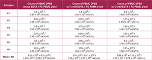
A statistically significant increase was observed only between the DPBS-alone group and the EDTA-supplemented groups (p=0.0015 and p=0.0044, respectively; Table 1). The higher EDTA concentration also increased PBMC counts; however, this difference was not statistically significant (p=0.8376; Table 1).
Comparison of Two Magnetic Bead-Based Cell Separation Platforms
We evaluated two magnetic bead–based cell separation platforms, by comparing standard outcome parameters, including post-separation cell viability and recovery yield. In addition, relative enrichment efficiency was assessed by downstream analysis of CD14 mRNA expression. All samples were processed using identical starting material (5 × 10⁶ PBMCs per separation; n=8 per group).
Overall cell viability, calculated as the percentage of viable output cells relative to the input, was comparable between the two systems (column-based system: 86.21% ± 8.58; column-free system: 85.60% ± 4.62; p > 0.05; Table 2). The number of recovered cells in the magnetically selected fraction was significantly higher with column-based system (4.1 × 10⁵ ± 0.3 × 10⁵) compared with column-free system (3.11 × 10⁵ ± 0.25 × 10⁵), indicating a statistically significant increase in recovered cell numbers with the column-based system (p < 0.001, Table 2).
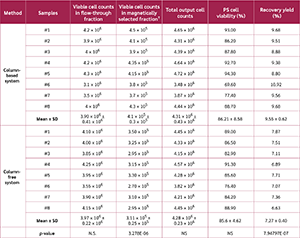
Accordingly, paired analyses demonstrated that recovery efficiency was significantly higher following column-based system (9.55 ± 0.62%) compared to column-free system (7.27% ± 0.40) (p<0.001, Table 2).
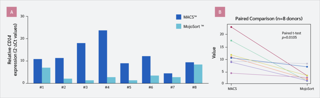
To further characterize the isolated fractions at the molecular level, CD14 gene expression was analyzed by quantitative RT-PCR (Figure 1). Following normalization to GAPDH, CD14 mRNA expression showed a 2.74 ± 2.11-fold increase in PBMCs, an 11.46 ± 5.83-fold increase in column-based system-selected fractions, and a 3.39 ± 2.64-fold increase in column-free system-selected fractions (Figure 1).
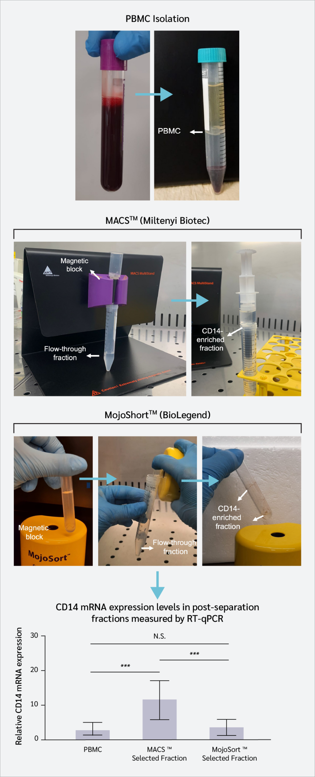

Cells obtained using the column-based system exhibited significantly higher CD14 mRNA expression compared to PBMCs and to fractions obtained using the column-free platform (p<0.001; Figure 1).
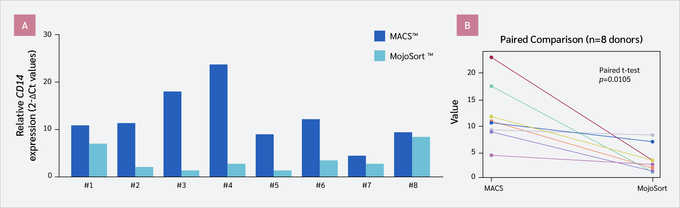
At the individual donor level, CD14 mRNA levels were consistently higher in fractions obtained using the column-based system than in those obtained using the column-free system, based on GAPDH-normalized values (2-ΔCt; Figure 2a). Paired statistical analysis (n=8) confirmed this observation, demonstrating a significant difference between the two separation methods (p=0.0105; Figure 2b), independent of donor variability.
Microscopic observations during trypan blue–based cell counting revealed differences in cellular composition between the two systems. Column-based system-derived fractions appeared more uniform, whereas Column-free system-derived fractions contained a higher proportion of additional cellular elements, including platelets (Figure S1d, Figure S1e). These observations, however, were qualitative and not quantitatively assessed.
To evaluate functional potential, magnetically selected fractions were subjected to in vitro differentiation for 7 days. Cells derived from both platforms exhibited morphology consistent with macrophage-like cells (Figure 3a, Figure 3b).
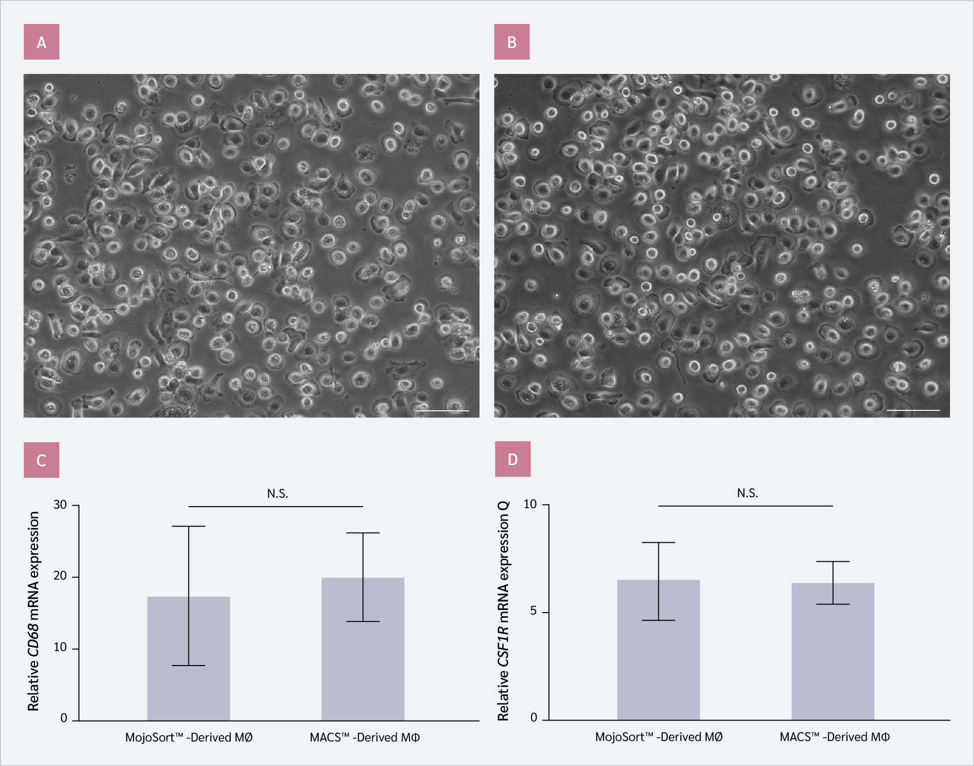
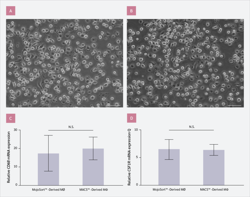
Expression of the macrophage-associated markers CD68 and colony-stimulating factor 1 receptor (CSF1R) was assessed by RT-qPCR. No statistically significant differences were observed between the two groups (p>0.05; Figure 3c, Figure 3d). CD68 mRNA expression increased by 6.51 ± 1.8-fold in column-free system-derived cells and by 6.4 ± 0.97-fold in column-based system-derived cells relative to their respective undifferentiated counterparts (Figure 3c). Similarly, CSF1R mRNA expression increased by 17.08 ± 9.77-fold and 19.82 ± 6.3-fold in column-free and column-based system-derived cells, respectively (Figure 3d).
Discussion
This study aimed to comparatively evaluate the performance of two commercially available magnetic bead–based cell separation platforms, under standardized experimental conditions. The comparison focused on cell recovery, viability, and enrichment efficiency as assessed by CD14 mRNA expression. Although CD14 is commonly associated with monocyte-lineage cells, it is important to note that the present study evaluates enrichment at the transcript level rather than at the phenotypic level.
MACS™, as one of the earliest and most widely adopted magnetic separation technologies, has been extensively used in previous studies (2,6,13). In the present work, both MACS™ and MojoSort™ systems were evaluated in parallel using identical starting material and processed by the same operator to minimize technical variability and ensure an unbiased comparison. In addition, we present practical optimization strategies for PBMC isolation and magnetic separation that may improve cell recovery and viability, particularly in settings with limited sample availability (Table S2).
Pre-analytical variables were found to play a critical role in downstream outcomes. The use of EDTA-containing blood collection tubes is recommended for RNA-based applications, as heparin may inhibit reverse transcriptase and DNA polymerase activity (14,15). However, EDTA may not be suitable for all cell types and applications (16), and its use should be considered in the context of the intended downstream analysis. Consistent with previous reports, the inclusion of EDTA in washing buffers reduced cell aggregation and improved recovery (12). Although Efthymiou et al. (12) reported no significant difference in PBMC counts between washing buffer with or without EDTA, flow cytometry analysis revealed higher frequencies of CD4+ and CD8+ T cells in the EDTA-supplemented group. In our experiments, phosphate-buffered saline (PBS) supplemented with EDTA increased PBMC yield by approximately 12%, which is particularly relevant when working with limited blood volumes (Table 1, Figure S1). Furthermore, timely processing of fresh blood samples and careful execution of density gradient centrifugation were essential for maintaining PBMC integrity and minimizing cell loss (17).
Magnetic separation workflows also require careful handling to ensure reproducibility. Preventing cell aggregation, optimizing column loading (for column-based system), and applying controlled washing steps were critical factors influencing recovery efficiency (1,2,7–9). The inclusion of BSA in separation buffers contributed to improved cell stability and reduced nonspecific binding, consistent with its known protective and blocking properties (18,19).
Post-separation viability is a key determinant of downstream usability. In the present study, both platforms demonstrated comparable viability (approximately 86%), with no statistically significant difference between systems. Although magnetic separation is generally associated with lower cell loss than fluorescence-activated cell sorting, some cell loss is expected due to mechanical handling and processing steps. Maintaining high viability after PBMC isolation and magnetic separation is critical for downstream applications, as mechanical stress and dead cells can promote nonspecific microbead binding. Although magnetic sorting is associated with approximately 10% cell loss (compared with approximately 70% for FACS) (20), our platforms showed similar overall losses with comparable post-sorting viability. Notably, although both systems showed similar overall recovery profiles, Column-based system yielded a significantly higher cell count in the magnetically selected fraction.
At the molecular level, CD14 mRNA expression assessed by RT-qPCR was used as an indirect indicator of enrichment efficiency in this study. Importantly, studies evaluating magnetic separation methods often report outcomes in terms of cell purity and phenotypic identity. However, when characterization is based solely on gene expression analysis, such as RT-qPCR, the results reflect transcript-level enrichment rather than direct evidence of protein expression or cellular composition. Therefore, careful interpretation of enrichment data is required, particularly in the absence of protein-level validation methods such as flow cytometry. In our study, column-based system achieved significantly higher CD14 mRNA-enriched cell yield, which aligns more closely with the expected ~10% monocyte fraction in peripheral blood (21). Importantly, these results reflect relative enrichment of CD14 mRNA expression rather than direct quantification of CD14 protein-positive cells. Therefore, interpretations regarding cell identity should be made with caution. The consistency of increased CD14 mRNA expression across all donors suggests that the observed differences are method-dependent rather than driven by inter-individual variability.
Functional assessment demonstrated that cells derived from both separation methods could undergo in vitro differentiation into macrophage-like cells, as evidenced by morphology and increased expression of macrophage-associated genes, including CD68 and CSF1R (Figure 3). No significant differences were observed between the two systems for these differentiation-associated markers, suggesting that both platforms generate cell fractions suitable for downstream functional applications. These findings are consistent with previous reports indicating that magnetic separation methods do not adversely affect subsequent differentiation capacity (21).
Qualitative microscopic observations revealed differences in cellular composition between fractions obtained with the two systems, with column-based system-derived fractions appearing more uniform, whereas column-free system-derived fractions contained additional cellular elements, such as platelets (Figure S1). However, these observations were not quantitatively assessed and should be interpreted cautiously, as no phenotypic validation was performed.
Both positive selection–based magnetic separation systems present inherent advantages and limitations. The column-based system employs a direct labeling strategy and column-based separation, which may contribute to higher retention efficiency of labeled cells. In contrast, the column-free system relies on indirect labeling and column-free separation, which may result in partial loss of labeled cells during handling steps. Moreover, negatively selected CD14-positive monocytes have been reported to be highly contaminated with platelets (3), whereas positive selection may be time-consuming (2). In our experimental setting, column-based system demonstrated higher recovery and stronger enrichment of CD14 mRNA expression, along with a shorter processing time. However, factors such as cost, required equipment, and workflow preferences may influence the choice of system in different laboratory settings.
A key limitation of this study is the absence of protein-level validation of the isolated cell fractions. CD14 expression was assessed exclusively at the mRNA level using RT-qPCR, and no flow cytometric or immunophenotypic analysis was performed. Consequently, the proportion of CD14 protein-positive cells within the isolated fractions and the precise cellular composition remain undetermined. The results should therefore be interpreted as measures of relative enrichment rather than phenotypic purity or definitive identification of monocytes. Future studies incorporating multiparametric flow cytometry and additional lineage markers will be necessary to confirm cellular identity and quantify population composition.
Despite the relatively small sample size, the use of a paired donor-matched design strengthened the statistical power and reduced the impact of inter-individual variability. All samples were processed in parallel under identical conditions, supporting the robustness and reproducibility of the comparative analysis. Nevertheless, validation in larger and independent cohorts will be important to confirm the generalizability of these findings.
Conclusion
This study provides a systematic comparison of two magnetic bead–based separation systems under controlled conditions and highlights key procedural factors that influence experimental outcomes. Both magnetic-bead cell sorting systems yielded viable cell fractions suitable for downstream molecular and functional analyses. However, column-based system demonstrated higher cell recovery and stronger enrichment of CD14 mRNA expression under the conditions tested. These findings support the use of column-based system as an efficient approach for generating CD14 mRNA-enriched cell fractions, while emphasizing that conclusions are limited to transcript-level enrichment in the absence of phenotypic validation.
Ethical Approval
The study was approved by the Kocaeli University Human Research Ethics Committee on July 18, 2024, with decision no. GOKAEK-2024/11.37.
Informed Consent
Written informed consent was obtained from parents or legal guardians of all participants.
Peer-review
Externally peer-reviewed
Author Contributions
Concept – A.B.; Design – A.B; Supervision – H.E.S, A.K.; Fundings – H.E.S., A.K.; Materials – A.B.; Data Collection and/or Processing – A.B., B.S., A.K.; Analysis and/or Interpretation – A.B.; Literature Review – A.B., B.S.; Writer – A.B., H.E.S., B.S.; Critical Reviews – A.K.
Conflict of Interest
The authors declare no conflict of interest.
Financial Disclosure
This study was supported by The Scientific and Technological Research Council of Türkiye under the Scientific and Technological Research Projects Funding Program (Grant No. 124R020).
AI Statement
During the preparation of this manuscript, the authors used OpenAI’s ChatGPT to improve text flow and Grammarly to enhance grammatical clarity. The authors critically reviewed and edited the content and take full responsibility for the accuracy and integrity of the manuscript.
Acknowledgment
We sincerely thank Prof. Esra Çağavi for generously providing partial support for the MACS™ system consumables used in this study and Assist. Prof. Hüseyin Uzuner and Sena Nur Bütünay for their valuable technical and experimental assistance.
References
Koc A, Akdeniz C, Cagavi E. Human macrophages directly modulate iPSC-derived cardiomyocytes at healthy state and congenital arrhythmia model in vitro. Pflugers Arch. 2022;474(12):1295–310. [CrossRef]
Mayer A, Lee S, Lendlein A, Jung F, Hiebl B. Efficacy of CD14+ blood monocytes/macrophages isolation: positive versus negative MACS protocol. Clin Hemorheol Microcirc. 2011;48(1):57–63. [CrossRef]
Nielsen MC, Andersen MN, Møller HJ. Monocyte isolation techniques significantly impact the phenotype of both isolated monocytes and derived macrophages in vitro. Immunology. 2020;159(1):63–74. [CrossRef]
Ohradanova-Repic A, Machacek C, Fischer MB, Stockinger H. Differentiation of human monocytes and derived subsets of macrophages and dendritic cells by the HLDA10 monoclonal antibody panel. Clin Transl Immunology. 2016;5(1):e55. [CrossRef]
Zarif JC, Hernandez JR, Verdone JE, Campbell SP, Drake CG, Pienta KJ. A phased strategy to differentiate human CD14+monocytes into classically and alternatively activated macrophages and dendritic cells. Biotechniques. 2016;61(1):33–41. [CrossRef]
Schmitz B, Radbruch A, Kümmel T, Wickenhauser C, Korb H, Hansmann ML, et al. Magnetic activated cell sorting (MACS)--a new immunomagnetic method for megakaryocytic cell isolation: comparison of different separation techniques. Eur J Haematol. 1994;52(5):267–75. [CrossRef]
Kalinina O, Minter LM, Sperling AI, Hollinger MK, Le P, Osborne BA, et al. Exopolysaccharide-treated dendritic cells effectively ameliorate acute graft-versus-host disease. Transplant Cell Ther. 2024;30(1):79.e1–e10. [CrossRef]
Damara A, Wegner J, Trzeciak ER, Kolb A, Nastaranpour M, Khatri R, et al. LL37/self-DNA complexes mediate monocyte reprogramming. Clin Immunol. 2024;265:110287. [CrossRef]
Xiao R, Zeng J, Bressler EM, Lu W, Grinstaff MW. Synthesis of bioactive (1→6)-β-glucose branched poly-amido-saccharides that stimulate and induce M1 polarization in macrophages. Nat Commun. 2022;13(1):4661. [CrossRef]
Livak KJ, Schmittgen TD. Analysis of relative gene expression data using real-time quantitative PCR and the 2(-Delta Delta C(T)) Method. Methods. 2001;25(4):402–8. [CrossRef]
Dinh B, Hoeksema MA, Spann NJ, Rendler J, Cobo I, Glass CK, et al. Isolation and cryopreservation of highly viable human peripheral blood mononuclear cells from whole blood: a guide for beginners. J Vis Exp. 2024;(212). [CrossRef]
Efthymiou A, Mureanu N, Pemberton R, Tai-MacArthur S, Mastronicola D, Scottà C, et al. Isolation and freezing of human peripheral blood mononuclear cells from pregnant patients. STAR Protoc. 2022;3(1):101204. [CrossRef]
Miltenyi S, Müller W, Weichel W, Radbruch A. High gradient magnetic cell separation with MACS. Cytometry. 1990;11(2):231–8. [CrossRef]
Ding M, Bullotta A, Caruso L, Gupta P, Rinaldo CR, Chen Y. An optimized sensitive method for quantitation of DNA/RNA viruses in heparinized and cryopreserved plasma. J Virol Methods. 2011;176(1-2):1–8. [CrossRef]
Marteau JB, Mohr S, Pfister M, Visvikis-Siest S. Collection and storage of human blood cells for mRNA expression profiling: a 15-month stability study. Clin Chem. 2005;51(7):1250–2. [CrossRef]
Betsou F, Gaignaux A, Ammerlaan W, Norris PJ, Stone M. Biospecimen science of blood for peripheral blood mononuclear cell (PBMC) functional applications. Curr Pathobiol Rep. 2019;7(2):17–27. [CrossRef]
Lehle S, Völkl S, Seitz K, Goossens C, Emons J, Ruebner M, et al. Effect of delayed isolation of peripheral blood mononuclear cells on cell viability and functionality. BMC Immunol. 2025;26(1):21. [CrossRef]
Mahmoud NN, Hammad AS, Al Kaabi AS, Alawi HH, Khatoon S, Al-Asmakh M. Evaluating the effects of BSA-coated gold nanorods on cell migration potential and inflammatory mediators in human dermal fibroblasts. J Funct Biomater. 2024;15(10):284. [CrossRef]
Hermanson GT. Homobifunctional crosslinkers. In: Hermanson GT, editor. Bioconjugate techniques. 3rd ed. Amsterdam: Elsevier; 2013. p. 275–98. [CrossRef]
Sutermaster BA, Darling EM. Considerations for high-yield, high-throughput cell enrichment: fluorescence versus magnetic sorting. Sci Rep. 2019;9(1):227. [CrossRef]
Weiss R, Gerdes W, Leonhardt F, Berthold R, Sack U, Grahnert A. A comparative study of two separation methods to isolate monocytes. Cytometry A. 2019;95(2):234–41. [CrossRef]
VOLUME
,
ISSUE
Correspondence
Received
Accepted
Published
Suggested Citation
DOI
License
