Immunogenic Potential of Foot and Mouth Virus Antigen O-146S Adjuvanted with Water-in-Oil and Water-in-Oil-in-Water Emulsion in Mice
Abstract
Objective:
Foot-and-mouth disease virus (FMDV) is a highly contagious viral pathogen that significantly impacts livestock health and productivity. This study aimed to evaluate the immunological responses to FMDV O-146S antigen adjuvanted with water-in-oil (W/O) and water-in-oil-in-water (W/O/W) emulsion in mice.
Materials and Methods:
Mice were immunized intramuscularly with a vaccine formulation containing the FMDV O-146S antigen combined with oil emulsion adjuvants at a predetermined ratio and then monitored for 21 days.
Results:
Compared to FMDV O-146S antigen alone, formulations containing oil emulsion adjuvants induced earlier and increased levels of anti-FMDV specific antibody responses, as demonstrated by elevated levels of anti-FMDV IgG (p<0.0001), IgG1 (p<0.0002), and IgG2a (p<0.0018). The increased IgG1 and IgG2a antibody isotype responses suggest activation of both Th2- and Th1-type immunity, respectively. Vaccines were also more effective at eliciting a cellular immune response, as measured by the effector T cells represented by CD3+CD8+ T cells (p<0.0001), IFN-γ secreting CD8+ T cells (p<0.0001), IFN-γ secreting CD3+ T cells (p<0.0001) and proliferation of splenocytes represented by higher stimulation index (p<0.0001), as well as the production of IFN-γ (p<0.0001), IL-4 (p<0.0002), and IL-1β (p<0.0006) cytokines.
Conclusion:
Oil emulsion adjuvants containing FMDV O-146S antigen significantly boost humoral and cell-mediated immune responses to the FMDV O-146S antigen in mice.
Keywords:
Foot, and, mouth, disease oil, emulsion, adjuvant vaccine immune, responseIntroduction
Foot-and-mouth disease (FMD) is a highly contagious and potentially fatal animal disease, according to the World Organization for Animal Health (WOAH), that primarily affects cloven-hoofed animals (1-3). The causative agent, foot-and-mouth disease virus (FMDV), is a member of the Picornaviridae family and belongs to the genus Aphthovirus. The symptoms of the disease include a sudden onset of fever and lesions on the mouth and the feet (4,5). The spread of FMD poses a significant risk to livestock, the agricultural sector, and the broader economy (6).
It is common practice to test FMDV vaccinations on susceptible pigs, cattle, and sheep for different types of vaccines. Comparatively, a mouse model can be used as a simple and inexpensive transitional model to test the immunological impact of a vaccine without the need for the extensive time, resources, and effort required by other animal models. Rodents, including water rats, moles, and brown house rats, have been shown to be susceptible to FMDV infection (7).
Water-in-oil (W/O) emulsion platforms are widely used in vaccine development due to their ability to enhance antigen presentation by modulating delivery to antigen-presenting cells (APCs) or through direct stimulation of immune cells (8). Commercially available adjuvants for FMDV vaccines include the mineral oil-based formulations such as Montanide ISA-206 and SA-201, as well as aluminum hydroxide. Oil emulsions are highly immunogenic because of their high reactogenicity (9).
This study aimed to evaluate, for the first time in mice, the immunogenic potential of novel W/O and water-in-oil-in-water (W/O/W) emulsion adjuvants developed by Coral Biotechnology, Inc. against the FMDV O-146S antigen.
Materials and Methods
Adjuvants
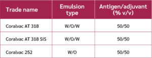
The emulsion adjuvants used in this study were Coralvac AT 318, Coralvac AT 318 SIS, and Coralvac 252. These adjuvants, listed in Table 1, were obtained from Coral Biotechnology, Inc. (Kartepe, Kocaeli, Türkiye).
FMDV O-146S antigen
Following approval from the Ministry of Agriculture and Forestry and the Foot-and-Mouth Disease Research Institute (Ankara, Türkiye) under permit number E-71037622-799-5127956, purified and sterilized FMDV O-146S antigen was obtained for this study. The antigen was supplied at a concentration of 110 μg/mL. Particles of the O-146S antigen, derived from FMDV strains, were used as the immunogen. The baby hamster kidney cell line (BHK21) was used for virus propagation.
Preparation of Emulsion Adjuvant-Based FMDV O-146S Antigen-Containing Vaccine Formulations
Vaccine formulations were prepared by mixing 3 µg/mL of FMDV O-146S antigen with each adjuvant (Coralvac AT 318, Coralvac AT 318 SIS, or Coralvac 252) in a 50:50 ratio to obtain a final volume of 15 mL. The adjuvants were filtered through 0.22 µm sterile filters prior to use in the vaccine formulation.
To prepare the formulations, the antigen phase was first transferred to a beaker and stirred at 100 rpm using a mechanical mixer. While stirring, the adjuvant was slowly added to the antigen phase. For Coralvac AT 318, the stirring speed was increased to 400 rpm and continued for 30 minutes. For Coralvac 252, the speed was also increased to 400 rpm, and the mixture was stirred for 2 minutes before being transferred to a homogenizer, where it was homogenized at 15,000 rpm for 10 minutes.
The prepared vaccine formulations were stored at +4°C for 24 hours.
Physical Stability Test
This experimental setup was designed to assess the stability of formulations under varying temperature conditions. Each antigen-adjuvant formulation was placed in a 1 mL sterile Eppendorf tube and incubated at both 25°C and +4°C to compare the stability of W/O and W/O/W emulsion-based vaccines. Following incubation periods of seven days, two months, and six months under both conditions, the tubes were evaluated for phase separation and visual agglomeration to determine the physical stability of the formulations over time.
According to characterization data provided by the manufacturer, the particle size distribution of the new adjuvant formulations was determined using integrated light scattering with a Malvern Pro Red instrument. Measurements of particle diameter range, specific surface area, and surface-volume mean diameter were conducted at room temperature immediately after emulsion preparation and again after six months of storage. The volume fraction of oil in the diluted emulsions was approximately 1:1000 in all cases.
Experimental Mice
All in vivo experiments were approved by the Ege University Animal Experiments Local Ethics Committee, with the decision numbered 2022-013 and dated January 25, 2023. Swiss albino mice, weighing 25–33 g, aged 4–6 weeks, were obtained from the Bornova Veterinary Control Research Institute. All mice were sexually mature at the time of acquisition. Prior to the experiments, the animals were acclimatized and quarantined at the Ege University Laboratory Animals Application and Research Center.
Immunological Studies
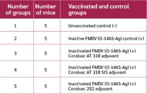
For this study, 25 Swiss albino mice (25–33 g, 4–6 weeks old) were divided into five groups (Table 2). Each mouse received a single intramuscular injection of 0.1 mL of FMDV O-146S antigen, either alone or formulated with W/O or W/O/W emulsion adjuvant. Vaccinations were administered into the hindlimb muscles on day zero. On day 21, the mice were euthanized; the injection sites were examined for local reactions, and blood and spleen samples were collected for immunological evaluation.
Detection of FMDV (O-146S-Ag)-Specific Antibody Response Using Indirect Enzyme-Linked Immunosorbent Assay (ELISA)
The levels of specific antibodies against the FMDV O-146S antigen in serum samples were determined using an indirect enzyme-linked immunosorbent assay (ELISA), as previously described (11). Briefly, microplates were coated with FMDV O-146S antigen at a concentration of 5 µg/mL in 100 µL per well. Serum samples were diluted 1:200, and 100 µL of the diluted sera was added to each well.
Cytokine Analysis by ELISA
Serum samples from each group were analyzed for cytokine levels using commercial ELISA kits (FineTest, China), according to the manufacturer's instructions. The detection range of IL-4, IL-1β and IFN-γ was IL-4 (15.625-1000 pg/ml; catalog no. EM0119), IL-1β (12.5-800 pg/ml; catalog no. EM0109), and IFN-γ (31.25-2000 pg/ml; catalog no. EM0093), respectively.
Splenic Cell Proliferation Assay
The splenic cell proliferation assay was performed as previously described (11). For each vaccination group, spleen cells were divided into three subgroups: a non-stimulated control group, an FMDV-stimulated group (O-146S antigen), and a lipopolysaccharide (LPS)-stimulated group (12).
Mouse spleens were aseptically excised and mechanically disrupted using a sterile mesh in Roswell Park Memorial Institute (RPMI)-1640 medium (Gibco, USA) supplemented with 10% bovine serum (Gibco, USA) and 1% penicillin-streptomycin (Gibco, USA) to obtain a homogeneous cellular suspension (13). The cell suspension was centrifuged at 450 g for 5 minutes and washed three times with RPMI-1640 medium. Cells were then resuspended in 7 mL of fresh medium and counted using a hemocytometer. A total of 5 × 10⁷ cells/mL were seeded into 96-well plates (100 µL/well). Plates were incubated at 37°C in a humidified atmosphere with 5% CO₂.
After 72 hours, splenocyte proliferation was assessed using the 3-(4,5-dimethylthiazol-2-yl)-2,5-diphenyltetrazolium bromide (MTT) assay. Mitochondrial dehydrogenase enzymes in proliferating cells convert MTT into a purple, water-insoluble formazan crystal, which is observed under an inverted microscope (14). The stimulation index (SI) was calculated using the following formula:
Stimulation index = (OD of stimulated cells − OD of unstimulated cells) / (OD of unstimulated cells)
Analysis of Cell-Mediated Immunity using Flow Cytometry
Using flow cytometry, the ratio of IFN-γ secreting CD3+CD8+ cells was determined. To achieve this goal, 3 μg/mL of FMDV O-146S antigen was added to the wells. The microtiter plates were then incubated in an incubator at 37°C and 5% CO₂ for 72 hours (19). After incubation, the samples in the microplates were transferred one by one to Eppendorf tubes. The samples were centrifuged at 2000 rpm for 5 minutes, and the supernatant was removed.
After centrifugation, 50 µL of fluorescein isothiocyanate (FITC)-conjugated rat anti-mouse CD3 molecular complex (BD Biosciences, USA), 50 µL of PerCP-conjugated rat anti-mouse CD8a (BD Biosciences, USA), 100 µL of fixation/permeabilization solution, and 50 µL of PE rat anti-mouse INF-γ (BD Biosciences, USA) antibodies were added to the Eppendorf tubes, respectively. Antibody working solutions were prepared separately by diluting 1 μL of anti-mouse CD3 antibody, 2.5 μL of anti-mouse CD8a antibody (in 3% fetal bovine serum [FBS]), and 2.5 μL of anti-mouse IFN-γ antibody in 1000 μL of phosphate-buffered saline (PBS) containing 3% FBS.
The samples were incubated at +4°C for 30 minutes in the dark at each stage and then centrifuged at 2000 rpm for 5 minutes between incubations. They were washed repeatedly twice with 250 µL of PBS containing 3% FBS. In the final stage, all samples were washed twice with 250 µL of washing solution, suspended in 300 µL, and analyzed in a flow cytometer device. Data analysis was conducted with BD Accuri™ C6 Software (BD Biosciences, USA).
Data Analysis
All data analyses and graphical presentations were conducted using GraphPad Prism 8 (GraphPad Software Inc., La Jolla, CA, USA). Statistical differences between groups were evaluated using analysis of variance (ANOVA) followed by post hoc testing. A p-value of <0.05 was considered statistically significant. Statistical significance was expressed as follows: n.s., not significant p≥0.05; p<0.05, significant.
Results
Stability of Prepared Oil Emulsion Adjuvant-Antigen Combinations
To evaluate the stability of the W/O and W/O/W emulsion formulations, each antigen-adjuvant combination intended for vaccination (FMDV O-146S antigen with Coralvac AT 318, Coralvac AT 318 SIS, and Coralvac 252) was incubated at two different temperatures.
At 25°C, none of the formulations showed visible agglomeration or phase separation after seven days. Following two months of incubation at this temperature, all formulations remained stable, except those containing Coralvac AT 318 and Coralvac AT 318 SIS, which exhibited signs of phase separation. After six months at 25°C, visual inspection revealed phase separation in all formulations (Figure S3).
At +4°C, all vaccine formulations were stable after seven days and two months. After six months of incubation at this temperature, phase separation was observed in all formulations, except for FMDV O-146S antigen combined with Coralvac AT 318 or Coralvac AT 318 SIS, which remained stable throughout the entire six-month storage period (Figure S4).
Particle size distribution of the emulsions was analyzed using integrated light scattering. Figures S1 and S2 display the particle size distributions immediately after preparation and after one year of storage. The resulting S-shaped curves indicate average droplet sizes ranging from 215.7 nm for Coralvac AT 318 to 249.2 nm for Coralvac 252 (Tables S1 and S2).
These findings suggest that +4°C was the ideal storage temperature for all formulations for a minimum of two months.
Determination of Specific Antibody Levels by Indirect ELISA
Twenty-one days after vaccination, serum samples were collected, and total immunoglobulin G (IgG) subclass antibody responses specific to the FMDV O-146S antigen were measured using indirect ELISA. Compared to the control group, mice that received the FMDV O-146S antigen formulated with adjuvants exhibited significantly higher absorbance values, indicating the development of an antigen-specific immune response.
As shown in Figure 1, all oil emulsion adjuvant formulations enhanced the production of IgG, IgG1, and IgG2a antibod-ies compared to the control group receiving antigen alone. Mice immunized with adjuvanted vac-cines displayed considerably higher IgG titers (****, p<0.0001). Specifically, IgG1 (associated with Th2-type responses) titers were increased in all adjuvant groups: FMDV O-146S antigen + Coralvac AT 318 (****, p<0.0001); FMDV O-146S antigen + Coralvac AT 318 SIS (***, p< 0.0002 ); and FMDV O-146S antigen + Coralvac 252 (***, p<0.0005 ).
Similarly, IgG2a antibody levels (indicative of Th1-type responses) were significantly higher in all adjuvanted groups compared to the control group: FMDV O-146S antigen + Coralvac AT 318 (p<0.0002); FMDV O-146S antigen + Coralvac AT 318 SIS (p<0.0004 ); and FMDV O-146S antigen + Coralvac 252 (p<0.0018) (Table 3 and Figure 1).
Determination of Cytokine Levels Following Vaccination with FMDV Formulation Groups
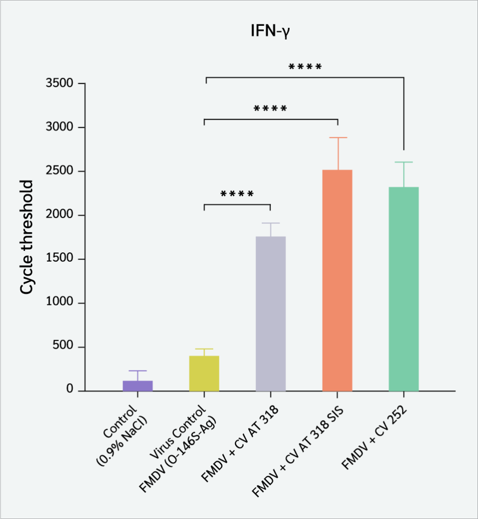
To evaluate the effects of each formulation on cytokine levels, serum samples from immunized mice were analyzed for Th1, Th2, and macrophage-associated cytokines. The higher levels of IFN-γ were detected in the serum from the mice immunized with FMDV O-146S antigen + CV AT 318, FMDV O-146S antigen + CV AT 318 SIS, and FMDV O-146S antigen + CV 252 compared to those from the mice immunized with the antigen alone (p<0.0001) (Figure 2).
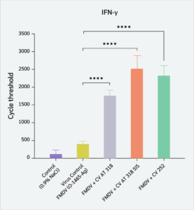
Figure 2:
Effects of different FMDV vaccine formulations on interferon-γ (IFN-γ) cytokine responses.
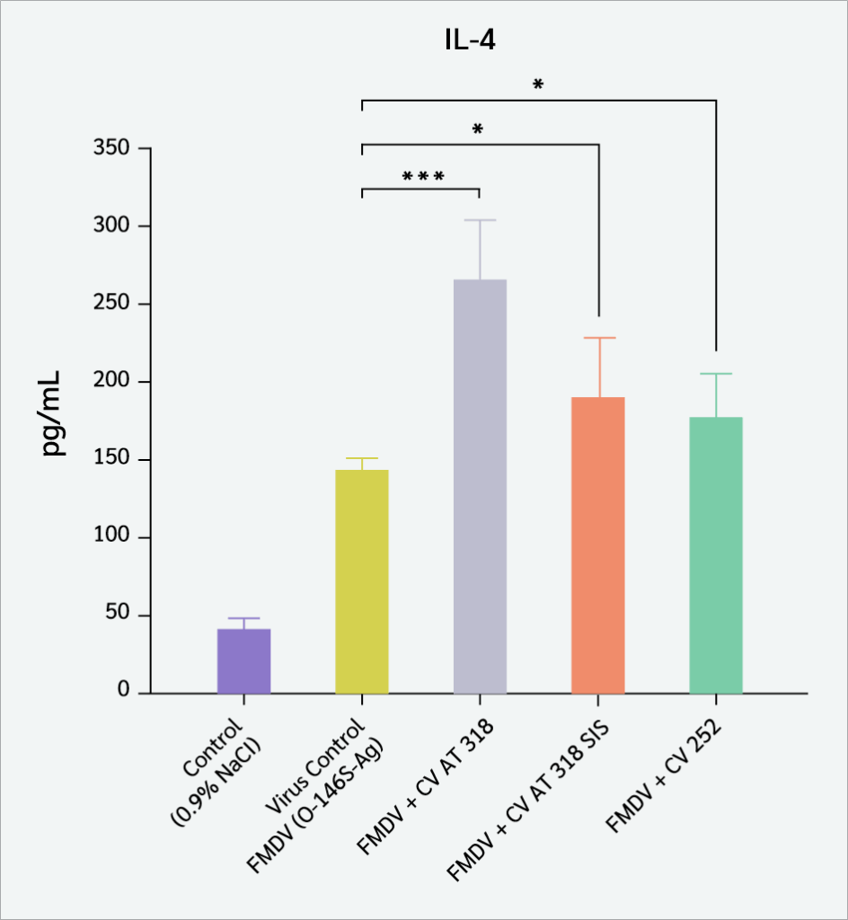
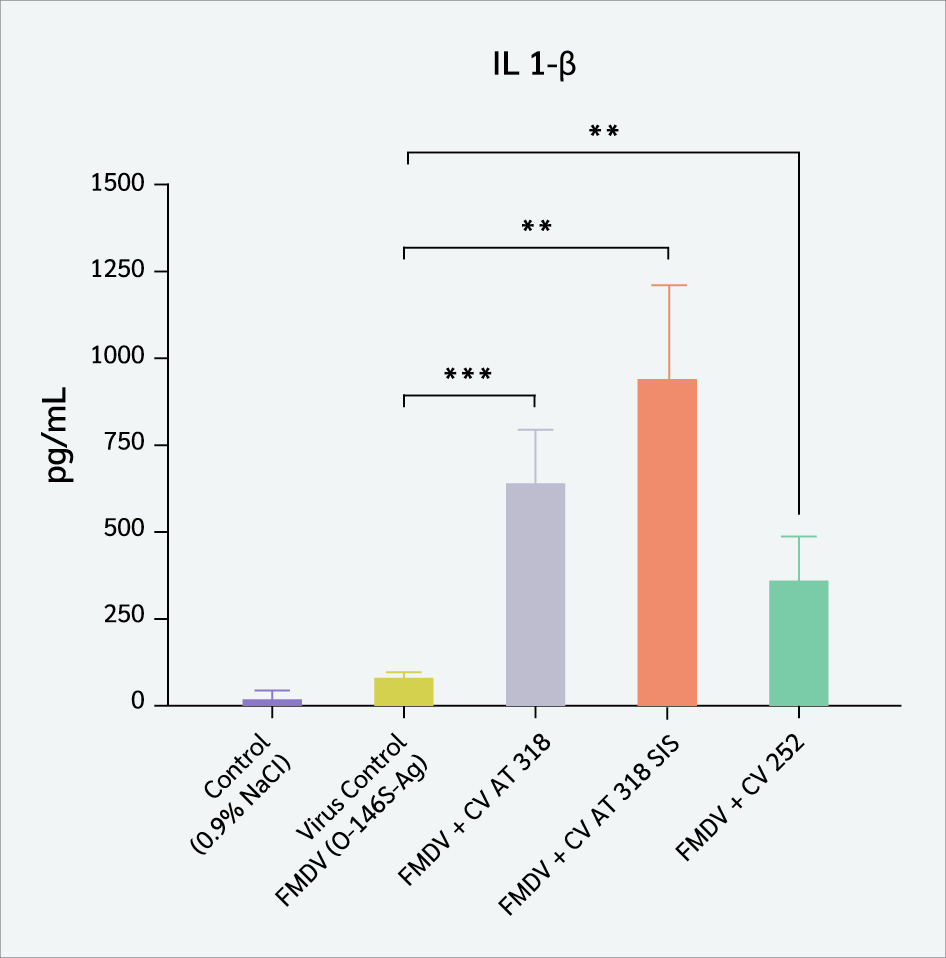
Serum from mice immunized with FMDV O-146S antigen + CV AT 318 represented higher levels of IL-4 in CD4+ T cells (p<0.0002) than serum from mice immunized with other adjuvanted groups; FMDV O-146S antigen + CV AT 318 SIS (p<0.033), and FMDV O-146S antigen + CV 252 (p<0.034) when compared with the control group (Figure 3). Serum from immunized mice showed statististically significantly increased levels of IL1-β in immune cells that produced it in the FMDV O-146S antigen + CV AT 318 (p<0.0006), FMDV O-146S antigen + CV AT 318 SIS (p<0.0011), and FMDV O-146S antigen + CV 252 (p<0.0053) groups compared to those from the mice vaccinated with the control group ( Figure 4).
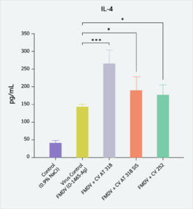
Figure 3:
Effects of different FMDV vaccine formulations on interleukin-4 (IL-4) cytokine responses.
These results demonstrate that the FMDV O-146S antigen, formulated with Coralvac adjuvants, effectively stimulated a balanced immune response by prompting both Th1 (IFN-γ) and Th2 (IL-4) cytokine production, as well as activating macrophage-mediated responses (IL-1β).
Evaluation of Splenocyte Proliferation in Immunized Mice
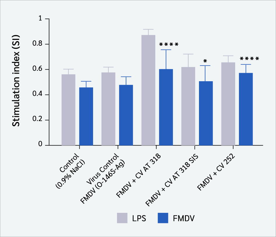
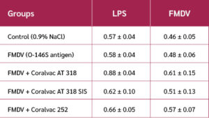
The MTT showed that the FMDV O-146S antigen + CV AT 318 (p<0.0001) and FMDV O-146S antigen + CV 252 (p<0.0001) groups exhibited significantly higher SI values and FMDV O-146S antigen + CV AT 318 SIS (p<0.049) compared to the group that received the FMDV O-146S antigen control alone (Table 4 and Figure 5)
Analysis of the Cellular Immune Response in Immunized Mice Using Flow Cytometry
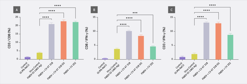
All vaccine formulation groups exhibited an increase in CD8+ T cell populations secreting IFN-γ after 72 hours of FMDV O-146S antigen stimulation. Notably, the CD3+CD8+ T cell immune response was significantly elevated in the FMDV O-146S antigen + CV AT 318, FMDV O-146S antigen + CV AT 318 SIS, and FMDV O-146S antigen + CV 252 groups compared to the control group (p<0.0001). The CD3+/CD8+ T cell ratio was approximately 20% across these three vaccine formulations (Figures 6a and S11).
The IFN-γ-secreting CD8+ T cells were found to be significantly higher when FMDV O-146S antigen + CV AT 318 (9.8%) vaccine combination was given (p<0.0001). In comparison to the control group, the FMDV O-146S antigen + CV AT 318 SIS group exhibited the second-highest response (7.9%) (p<0.0001), followed by the FMDV O-146S antigen + CV 252 group (4.4%) (p<0.0005) (Figures 6b and S12).
When the mice were injected the FMDV O-146S antigen + CV AT 318, the IFN-γ producing CD3+ T cell ratio was 12.7%, which was statistically significantly higher than the virus control group (p<0.0001). It was 12.5% in mice that were given FMDV o 146S antigen + AT 318 SIS (p<0.0001) compared to the control group. It was demonstrated that FMDV O-146S antigen + CV 252 had the third-highest proportion of IFN-γ-secreting T cells (8.4%) among all the groups and was significantly different from the control group (p<0.0001) (Figures 6c and S13).
Discussion
The results of this study showed that the newly developed oil emulsion adjuvants significantly enhanced adaptive immune responses and exhibited strong adjuvant properties when combined with the FMDV O-146S antigen-containing vaccine formulation in mice. Adding an adjuvant to a co-administered antigen enhances the immunogenicity of the substance by stimulating a more robust and durable immune response (16). The use of a mouse model for evaluating vaccines offers several advantages, primarily in terms of reducing cost and time by allowing the assessment of the immune response before testing them in the primary host. The pathogenesis of FMDV in mice is influenced by multiple factors, including viral characteristics, the animal strain, and the method of viral penetration (17). Results from studies on target species often balance results from mouse studies. Therefore, the mouse model can reliably predict the immune response to foot-and-mouth disease viruses in cattle and pigs (17,18). Furthermore, several studies have used mice to evaluate the effectiveness of vaccines in both mice and cattle, and the results have been similar for both species (16,19,20).
Emulsion adjuvant studies have demonstrated that antigens can adsorb at the oil-water interface through both hydrophobic and electrostatic forces, resulting in significant inter-protein interactions and conformational changes in the adsorbed protein (19,20). In the current study, visual inspection of phase separation variations was used to investigate the thermal stability of FMDV vaccines across various temperatures. Inactivated FMDV is relatively stable at +4°C, but it exhibits poor stability at temperatures above 37°C, consistent with findings from studies on the live virus (21). Problems with early in vitro characterization of the vaccine may lead to the loss of the effective antigens going undetected and, ultimately, to a failed immunization (22). As a result, there is an urgent need for an FMDV vaccination with greater stability of vaccine formulation. Figures S3 and S4 present the findings of this study, which examine the stability of FMDV O-146S antigen-containing vaccine formulations with new adjuvants over time. The results demonstrate that vaccines are more stable when stored at 25°C for seven days and at +4°C for two months.
Previous research from our group demonstrated that when the adjuvant was evaluated independently, it did not induce any adverse effects or elicit a reaction against itself (23). The induction of neutralizing antibodies is responsible for the protective immunity to the antigen. Vaccines are designed primarily to induce specific humoral immunity. Antibody induction is typically credited as the cause of protective immunity against FMDV (24). Therefore, the efficacy of an adjuvant depends on its ability to induce faster, stronger, and more sustained responses from the immune system in the form of neutralizing antibodies (25). Specifically, total IgG antibody titers and neutralizing antibody responses can be effectively induced against FMDV using newly designed oil emulsion adjuvants. Immunization-induced IgG subclass is a predictor for the balance between Th2- and Th1-type cytokines (26).
In general, IgG2a antibodies are associated with Th1 cells, while IgG1 antibodies are induced by Th2 cells (27,28). Both IgG1 and IgG2a may have potent neutralizing abilities; however, IgG2a is more likely to contribute to virus clearance due to its important interaction with complement components and Fc receptors (29). The effectiveness of FMD vaccinations can be estimated indirectly by analyzing serological reactions following vaccination, as humoral immunity is linked to clinical protection against FMD (30). The antibody isotype response may indicate the kind of immune activity (Th1 vs. Th2) occurring in vivo (31). In this study, IgG isotypes against the FMDV O-146S antigen were detected using an indirect ELISA method. Following a single intramuscular injection, mice immunized with oil adjuvant formulations containing inactivated FMDV produced FMDV O-146S antigen-specific IgG1 and IgG2a, indicating the stimulation of Th1 and Th2 cells, which will have a significant effect in providing good protection against FMD virus infection through viral clearance.
A higher lymphoproliferative response was observed in mice injected with the inactive FMDV O-146S antigen + CV AT 318, FMDV O-146S antigen + CV AT 318 SIS, and FMDV O-146S antigen + CV 252 vaccine groups, as determined by studies of virus-specific cellular response. These findings suggest that the use of these adjuvants enhances the adaptive immune response against the FMDV O-146S antigen. Immunizing mice with FMDV O-146S antigen has been demonstrated to stimulate T-cell responses and increase the number of CD8+ T cells in the spleen (32). In addition, oil emulsion formulations of FMDV cause FMDV O-146S antigen-specific CD8+ T cells to proliferate and secrete IFN-γ. It is generally known that IFN-γ plays a role in switching the isotype of immunoglobulins, which ultimately results in a greater number of IgG2a types (33). This result is consistent with the high amounts of IgG2a that were acquired, as well as the proliferation levels that were found in the FMDV + CV AT 318, FMDV + CV AT 318 SIS, and FMDV O-146S antigen + CV 252 groups. These findings suggest that FMDV O-146S antigen adjuvanted with newly designed oil emulsion adjuvants elicits a significant cellular response.
Conclusion
The results of this study reveal that both humoral and cell-mediated immune responses against FMDV O-146S antigen can be developed in mice given a vaccine containing FMDV O-146S antigen formulated with novel W/O and W/O/W type emulsion adjuvants.
Ethical Approval
All in vivo experiments were approved by the Ege University Animal Experiments Local Ethics Committee, with the decision numbered 2022-013 and dated January 25, 2023.
Informed Consent
N.A.
Peer-review
Externally peer-reviewed
Author Contributions
Concept – A.N.; Design – A.N.; Supervision – A.N.; Fundings – A.N.; Materials – A.N.; Data Collection and/or Processing – A.N., K.A.M.H.A.; Analysis and/or Interpretation – A.N., K.A.M.H.A.; Literature Review – A.N., K.A.M.H.A.; Writer – A.N., K.A.M.H.A.; Critical Reviews – A.N.
Conflict of Interest
The authors declare no conflict of interest.
Financial Disclosure
The authors declared that this study has received no financial support.
References
Lin X, Yang Y, Song Y, Li S, Zhang X, Su Z, et al. Possible action of transition divalent metal ions at the inter-pentameric interface of inactivated foot-and-mouth disease virus provide a simple but effective approach to enhance stability. J Virol. 2021;95(7):e02431-20. [CrossRef]
Gao H, Ma J. Spatial distribution and risk areas of foot and mouth disease in mainland China. Prev Vet Med. 2021;189:105311. [CrossRef]
Ren HR, Li MT, Wang YM, Jin Z, Zhang J. The risk factor assessment of the spread of foot-and-mouth disease in mainland China. J Theor Biol. 2021;512:110558. [CrossRef]
Arzt J, Belsham GJ, Lohse L, Bøtner A, Stenfeldt C. Transmission of foot-and-mouth disease from persistently infected carrier cattle to naive cattle via transfer of oropharyngeal fluid. mSphere. 2018;3(5):e00365-18. [CrossRef]
Maree F, de Klerk-Lorist LM, Gubbins S, Zhang F, Seago J, Pérez-Martín E, et al. Differential persistence of foot-and-mouth disease virus in African buffalo is related to virus virulence. J Virol. 2016;90(10):5132-40. [CrossRef]
Yamada M, Fukai K, Morioka K, Nishi T, Yamazoe R, Kitano R, et al. Early pathogenesis of the foot-and-mouth disease virus O/JPN/2010 in experimentally infected pigs. J Vet Med Sci. 2018;80(4):689-700. [CrossRef]
Chen Y, Hu Y, Chen H, Li X, Qian P. A ferritin nanoparticle vaccine for foot-and-mouth disease virus elicited partial protection in mice. Vaccine. 2020;38(35):5647-52. [CrossRef]
Wang ZB, Xu J. Better adjuvants for better vaccines: progress in adjuvant delivery systems, modifications, and adjuvant-antigen codelivery. Vaccines (Basel). 2020;8(1):128. [CrossRef]
Chwalek M, Lalun N, Bobichon H, Plé K, Voutquenne-Nazabadioko L. Structure-activity relationships of some hederagenin diglycosides: haemolysis, cytotoxicity and apoptosis induction. Biochim Biophys Acta. 2006;1760(9):1418-27. [CrossRef]
Wang D, Cao H, Li J, Zhao B, Wang Y, Zhang A, et al. Adjuvanticity of aqueous extracts of Artemisia rupestris L. for inactivated foot-and-mouth disease vaccine in mice. Res Vet Sci. 2019;124:191-9. [CrossRef]
Alsakini KAMH, Çöven FO, Nalbantsoy A. Adjuvant effects of novel water/oil emulsion formulations on immune responses against infectious bronchitis (IB) vaccine in mice. Biologicals. 2024;85:101736. [CrossRef]
da Cruz Filho IJ, da Silva Barros BR, de Souza Aguiar LM, Navarro CDC, Ruas JS, de Lorena VMB, et al. Lignins isolated from Prickly pear cladodes of the species Opuntia fícus-indica (Linnaeus) Miller and Opuntia cochenillifera (Linnaeus) Miller induces mice splenocytes activation, proliferation and cytokines production. Int J Biol Macromol. 2019;123:1331-9. [CrossRef]
Nalbantsoy A, Nesil T, Erden S, Calış I, Bedir E. Adjuvant effects of Astragalus saponins Macrophyllosaponin B and Astragaloside VII. J Ethnopharmacol. 2011;134(3):897-903. [CrossRef]
Mosmann T. Rapid colorimetric assay for cellular growth and survival: application to proliferation and cytotoxicity assays. J Immunol Methods. 1983;65(1-2):55-63. [CrossRef]
Abu-Rish EY, Mansour AT, Mansour HT, Dahabiyeh LA, Aleidi SM, Bustanji Y. Pregabalin inhibits in vivo and in vitro cytokine secretion and attenuates spleen inflammation in lipopolysaccharide/concanavalin A -induced murine models of inflammation. Sci Rep. 2020;10(1):4007. [CrossRef]
Gnazzo V, Quattrocchi V, Soria I, Pereyra E, Langellotti C, Pedemonte A, et al. Mouse model as an efficacy test for foot-and-mouth disease vaccines. Transbound Emerg Dis. 2020;67(6):2507-20. [CrossRef]
Dus Santos MJ, Wigdorovitz A, Maradei E, Periolo O, Smitsaart E, Borca MV, et al. A comparison of methods for measuring the antibody response in mice and cattle following vaccination against foot and mouth disease. Vet Res Commun. 2000;24(4):261-73. [CrossRef]
Krisdiyantoro A, Rahmahani J, Puspitasari Y, Soeharsono S, Kurniawan Y, Azzahra F, et al. Evaluation of immune response in mice (Mus musculus) post-immunization with foot-and-mouth disease virus master seed candidate vaccine [dataset]. 2025. Available from: https://doi.org/10.6084/m9.figshare.28327520.v1
Fox CB, Kramer RM, Barnes V L, Dowling QM, Vedvick TS. Working together: interactions between vaccine antigens and adjuvants. Ther Adv Vaccines. 2013;1(1):7-20. [CrossRef]
Wei Y, Xiong J, Larson NR, Iyer V, Sanyal G, Joshi SB, et al. Effect of 2 emulsion-based adjuvants on the structure and thermal stability of Staphylococcus aureus alpha-toxin. J Pharm Sci. 2018;107(9):2325-34. [CrossRef]
Doel TR, Baccarini PJ. Thermal stability of foot-and-mouth disease virus. Arch Virol. 1981;70(1):21-32. [CrossRef]
Yang Y, Zhao Q, Li Z, Sun L, Ma G, Zhang S, Su Z. Stabilization study of inactivated foot and mouth disease virus vaccine by size-exclusion HPLC and differential scanning calorimetry. Vaccine. 2017;35(18):2413-9. [CrossRef]
Alsakini KAMH, Sanci E, Buhur A, Yavasoglu A, Karabay Yavasoglu NÜ, Nalbantsoy A. Single and repeat-dose toxicity and local tolerance assessment of newly developed oil emulsion adjuvant formulations for veterinary purposes. Drug Chem Toxicol. 2024;47(6):827-38. [CrossRef]
McCullough KC, De Simone F, Brocchi E, Capucci L, Crowther JR, Kihm U. Protective immune response against foot-and-mouth disease. J Virol. 1992;66(4):1835-40. [CrossRef]
Dar P, Kalaivanan R, Sied N, Mamo B, Kishore S, Suryanarayana VV, et al. Montanide ISA™ 201 adjuvanted FMD vaccine induces improved immune responses and protection in cattle. Vaccine. 2013;31(33):3327-32. [CrossRef]
Cribbs DH, Ghochikyan A, Vasilevko V, Tran M, Petrushina I, Sadzikava N, et al. Adjuvant-dependent modulation of Th1 and Th2 responses to immunization with beta-amyloid. Int Immunol. 2003;15(4):505-14. [CrossRef]
Cao W, Chen Y, Alkan S, Subramaniam A, Long F, Liu H, et al. Human T helper (Th) cell lineage commitment is not directly linked to the secretion of IFN-gamma or IL-4: characterization of Th cells isolated by FACS based on IFN-gamma and IL-4 secretion. Eur J Immunol. 2005;35(9):2709-17. [CrossRef]
Su B, Wang J, Wang X, Jin H, Zhao G, Ding Z, et al. The effects of IL-6 and TNF-alpha as molecular adjuvants on immune responses to FMDV and maturation of dendritic cells by DNA vaccination. Vaccine. 2008;26(40):5111-22. [CrossRef]
Quan FS, Vunnava A, Compans RW, Kang SM. Virus-like particle vaccine protects against 2009 H1N1 pandemic influenza virus in mice. PLoS One. 2010;5(2):e9161. [CrossRef]
Ahn YH, Chathuranga WAG, Shim YJ, Haluwana DK, Kim EH, Yoon IJ, et al. The potential adjuvanticity of CAvant®SOE for foot-and-mouth disease vaccine. Vaccines (Basel). 2021;9(10):1091. [CrossRef]
Oh Y, Fleming L, Statham B, Hamblin P, Barnett P, Paton DJ, et al. Interferon-γ induced by in vitro re-stimulation of CD4+ T-cells correlates with in vivo FMD vaccine induced protection of cattle against disease and persistent infection. PLoS One. 2012;7(9):e44365. [CrossRef]
Langellotti C, Quattrocchi V, Alvarez C, Ostrowski M, Gnazzo V, Zamorano P, et al. Foot-and-mouth disease virus causes a decrease in spleen dendritic cells and the early release of IFN-α in the plasma of mice. Differences between infectious and inactivated virus. Antiviral Res. 2012;94(1):62-71. [CrossRef]
Abbas AK, Lichtman AH, Pillai S. Cellular and molecular immunology. 7th ed. Philadelphia (PA): Elsevier/Saunders; 2012.
VOLUME
,
ISSUE
Correspondence
Received
Accepted
Published
Suggested Citation
DOI
License

